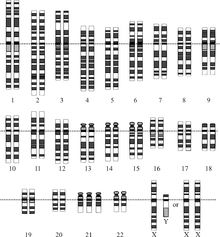
A chromosomal abnormality, chromosomal anomaly, chromosomal aberration, chromosomal mutation, or chromosomal disorder is a missing, extra, or irregular portion of chromosomal DNA. These can occur in the form of numerical abnormalities, where there is an atypical number of chromosomes, or as structural abnormalities, where one or more individual chromosomes are altered. Chromosome mutation was formerly used in a strict sense to mean a change in a chromosomal segment, involving more than one gene. Chromosome anomalies usually occur when there is an error in cell division following meiosis or mitosis. Chromosome abnormalities may be detected or confirmed by comparing an individual's karyotype, or full set of chromosomes, to a typical karyotype for the species via genetic testing.
Sometimes chromosomal abnormalities arise in the early stages of an embryo, sperm, or infant. They can be caused by various environmental factors. The implications of chromosomal abnormalities depend on the specific problem, they may have quite different ramifications. Some examples are Down syndrome and Turner syndrome.
Numerical abnormality

An abnormal number of chromosomes is known as aneuploidy, and occurs when an individual is either missing a chromosome from a pair (resulting in monosomy) or has more than two chromosomes of a pair (trisomy, tetrasomy, etc.). Aneuploidy can be full, involving a whole chromosome missing or added, or partial, where only part of a chromosome is missing or added. Aneuploidy can occur with sex chromosomes or autosomes.
Rather than having monosomy, or only one copy, the majority of aneuploid people have trisomy, or three copies of one chromosome. An example of trisomy in humans is Down syndrome, which is a developmental disorder caused by an extra copy of chromosome 21; the disorder is therefore also called trisomy 21.
An example of monosomy in humans is Turner syndrome, where the individual is born with only one sex chromosome, an X.
Sperm aneuploidy
Exposure of males to certain lifestyle, environmental and/or occupational hazards may increase the risk of aneuploid spermatozoa. In particular, risk of aneuploidy is increased by tobacco smoking, and occupational exposure to benzene, insecticides, and perfluorinated compounds. Increased aneuploidy is often associated with increased DNA damage in spermatozoa.
Structural abnormalities


When the chromosome's structure is altered, this can take several forms:
- Deletions: A portion of the chromosome is missing or has been deleted. Known disorders in humans include Wolf–Hirschhorn syndrome, which is caused by partial deletion of the short arm of chromosome 4; and Jacobsen syndrome, also called the terminal 11q deletion disorder.
- Duplications: A portion of the chromosome has been duplicated, resulting in extra genetic material. Known human disorders include Charcot–Marie–Tooth disease type 1A, which may be caused by duplication of the gene encoding peripheral myelin protein 22 (PMP22) on chromosome 17.
- Inversions: A portion of the chromosome has broken off, turned upside down, and reattached, therefore the genetic material is inverted.
- Insertions: A portion of one chromosome has been deleted from its normal place and inserted into another chromosome.
- Translocations: A portion of one chromosome has been transferred to another chromosome. There are two main types of translocations:
- Reciprocal translocation: Segments from two different chromosomes have been exchanged.
- Robertsonian translocation: An entire chromosome has attached to another at the centromere - in humans, these only occur with chromosomes 13, 14, 15, 21, and 22.
- Rings: A portion of a chromosome has broken off and formed a circle or ring. This happens with or without the loss of genetic material.
- Isochromosome: Formed by the mirror image copy of a chromosome segment including the centromere.
Chromosome instability syndromes are a group of disorders characterized by chromosomal instability and breakage. They often lead to an increased tendency to develop certain types of malignancies.
Inheritance
Most chromosome abnormalities occur as an accident in the egg cell or sperm, and therefore the anomaly is present in every cell of the body. Some anomalies, however, can happen after conception, resulting in Mosaicism (where some cells have the anomaly and some do not). Chromosome anomalies can be inherited from a parent or be "de novo". This is why chromosome studies are often performed on parents when a child is found to have an anomaly. If the parents do not possess the abnormality it was not initially inherited; however, it may be transmitted to subsequent generations.
Acquired chromosome abnormalities
Most cancers, if not all, could cause chromosome abnormalities, with either the formation of hybrid genes and fusion proteins, deregulation of genes and overexpression of proteins, or loss of tumor suppressor genes (see the "Mitelman Database" and the Atlas of Genetics and Cytogenetics in Oncology and Haematology,). Furthermore, certain consistent chromosomal abnormalities can turn normal cells into a leukemic cell such as the translocation of a gene, resulting in its inappropriate expression.
DNA damage during spermatogenesis
During the mitotic and meiotic cell divisions of mammalian gametogenesis, DNA repair is effective at removing DNA damages. However, in spermatogenesis the ability to repair DNA damages decreases substantially in the latter part of the process as haploid spermatids undergo major nuclear chromatin remodeling into highly compacted sperm nuclei. As reviewed by Marchetti et al., the last few weeks of sperm development before fertilization are highly susceptible to the accumulation of sperm DNA damage. Such sperm DNA damage can be transmitted unrepaired into the egg where it is subject to removal by the maternal repair machinery. However, errors in maternal DNA repair of sperm DNA damage can result in zygotes with chromosomal structural aberrations.
Melphalan is a bifunctional alkylating agent frequently used in chemotherapy. Meiotic inter-strand DNA damages caused by melphalan can escape paternal repair and cause chromosomal aberrations in the zygote by maternal misrepair. Thus both pre- and post-fertilization DNA repair appear to be important in avoiding chromosome abnormalities and assuring the genome integrity of the conceptus.
Detection
Depending on the information one wants to obtain, different techniques and samples are needed.
- For the prenatal diagnosis of a fetus, amniocentesis, chorionic villus sampling or circulating foetal cells would be collected and analysed in order to detect possible chromosomal abnormalities.
- For the preimplantational diagnosis of an embryo, a blastocyst biopsy would be performed.
- For a lymphoma or leukemia screening the technique used would be a bone marrow biopsy.
Nomenclature
-
 Three chromosomal abnormalities with ISCN nomenclature, with increasing complexity: (A) A tumour karyotype in a male with loss of the Y chromosome, (B) Prader–Willi Syndrome i.e. deletion in the 15q11-q12 region and (C) an arbitrary karyotype that involves a variety of autosomal and allosomal abnormalities.
-
 Human karyotype with annotated bands and sub-bands as used for the nomenclature of chromosome abnormalities. It shows dark and white regions as seen on G banding. Each row is vertically aligned at centromere level. It shows 22 homologous autosomal chromosome pairs, both the female (XX) and male (XY) versions of the two sex chromosomes, as well as the mitochondrial genome (at bottom left).
The International System for Human Cytogenomic Nomenclature (ISCN) is an international standard for human chromosome nomenclature, which includes band names, symbols and abbreviated terms used in the description of human chromosome and chromosome abnormalities. Abbreviations include a minus sign (-) for chromosome deletions, and del for deletions of parts of a chromosome.
See also
- Aneuploidy
- Chromosome segregation
- Genetic disorder
- Gene therapy
- Nondisjunction
- Obstetrical complications
References
- "Chromosomal Abnormalities", Understanding Genetics: A New York, Mid-Atlantic Guide for Patients and Health Professionals, Genetic Alliance, 2009-07-08, retrieved 2023-09-27
- NHGRI. 2006. Chromosome Abnormalities Archived 2006-09-25 at the Wayback Machine
- Rieger, R., Michaelis, A., Green, M.M. (1968). "Mutation". A glossary of genetics and cytogenetics: Classical and molecular. New York: Springer-Verlag. ISBN 978-0-387-07668-3.
- Chen H (2006). Atlas of genetic diagnosis and counseling. Totowa, N.J: Humana Press. ISBN 978-1-58829-681-8.
- ^ Gardner, R. J. M. (2012). Chromosome abnormalities and genetic counseling. Sutherland, Grant R., Shaffer, Lisa G. (4th ed.). Oxford: Oxford University Press. ISBN 978-0-19-974915-7. OCLC 769344040.
- "Numerical Abnormalities: Overview of Trisomies and Monosomies - Health Encyclopedia - University of Rochester Medical Center". www.urmc.rochester.edu. Retrieved 2020-11-17.
- Patterson D (2009-07-01). "Molecular genetic analysis of Down syndrome". Human Genetics. 126 (1): 195–214. doi:10.1007/s00439-009-0696-8. ISSN 1432-1203. PMID 19526251. S2CID 10403507.
- "Turner Syndrome". National Institute of Child Health and Human Development. Retrieved 2020-11-17.
- Templado C, Uroz L, Estop A (2013). "New insights on the origin and relevance of aneuploidy in human spermatozoa". Mol. Hum. Reprod. 19 (10): 634–43. doi:10.1093/molehr/gat039. PMID 23720770.
- Shi Q, Ko E, Barclay L, Hoang T, Rademaker A, Martin R (2001). "Cigarette smoking and aneuploidy in human sperm". Mol. Reprod. Dev. 59 (4): 417–21. doi:10.1002/mrd.1048. PMID 11468778. S2CID 35230655.
- Rubes J, Lowe X, Moore D, Perreault S, Slott V, Evenson D, Selevan SG, Wyrobek AJ (1998). "Smoking cigarettes is associated with increased sperm disomy in teenage men". Fertil. Steril. 70 (4): 715–23. doi:10.1016/S0015-0282(98)00261-1. PMID 9797104.
- Xing C, Marchetti F, Li G, Weldon RH, Kurtovich E, Young S, Schmid TE, Zhang L, Rappaport S, Waidyanatha S, Wyrobek AJ, Eskenazi B (2010). "Benzene exposure near the U.S. permissible limit is associated with sperm aneuploidy". Environ. Health Perspect. 118 (6): 833–9. Bibcode:2010EnvHP.118..833X. doi:10.1289/ehp.0901531. PMC 2898861. PMID 20418200.
- Xia Y, Bian Q, Xu L, Cheng S, Song L, Liu J, Wu W, Wang S, Wang X (2004). "Genotoxic effects on human spermatozoa among pesticide factory workers exposed to fenvalerate". Toxicology. 203 (1–3): 49–60. Bibcode:2004Toxgy.203...49X. doi:10.1016/j.tox.2004.05.018. PMID 15363581. S2CID 36073841.
- Xia Y, Cheng S, Bian Q, Xu L, Collins MD, Chang HC, Song L, Liu J, Wang S, Wang X (2005). "Genotoxic effects on spermatozoa of carbaryl-exposed workers". Toxicol. Sci. 85 (1): 615–23. doi:10.1093/toxsci/kfi066. PMID 15615886.
- Governini L, Guerranti C, De Leo V, Boschi L, Luddi A, Gori M, Orvieto R, Piomboni P (2014). "Chromosomal aneuploidies and DNA fragmentation of human spermatozoa from patients exposed to perfluorinated compounds". Andrologia. 47 (9): 1012–9. doi:10.1111/and.12371. hdl:11365/982323. PMID 25382683. S2CID 13484513.
- "Chromosome Abnormalities". atlasgeneticsoncology.org. Archived from the original on 14 August 2006. Retrieved 9 May 2018.
- "Chromosomes, Leukemias, Solid Tumors, Hereditary Cancers". atlasgeneticsoncology.org. Archived from the original on 28 January 2015. Retrieved 9 May 2018.
- "Mitelman Database of Chromosome Aberrations and Gene Fusions in Cancer". Archived from the original on 2016-05-29.
- "Atlas of Genetics and Cytogenetics in Oncology and Haematology". atlasgeneticsoncology.org. Archived from the original on 2011-02-23.
- Chaganti RS, Nanjangud G, Schmidt H, Teruya-Feldstein J (October 2000). "Recurring chromosomal abnormalities in non-Hodgkin's lymphoma: biologic and clinical significance". Seminars in Hematology. 37 (4): 396–411. doi:10.1016/s0037-1963(00)90019-2. ISSN 0037-1963. PMID 11071361.
- Baarends WM, van der Laan R, Grootegoed JA (2001). "DNA repair mechanisms and gametogenesis". Reproduction. 121 (1): 31–9. doi:10.1530/reprod/121.1.31. hdl:1765/9599. PMID 11226027.
- ^ Marchetti F, Bishop J, Gingerich J, Wyrobek AJ (2015). "Meiotic interstrand DNA damage escapes paternal repair and causes chromosomal aberrations in the zygote by maternal misrepair". Sci Rep. 5: 7689. Bibcode:2015NatSR...5.7689M. doi:10.1038/srep07689. PMC 4286742. PMID 25567288.
- Warrender JD, Moorman AV, Lord P (2019). "A fully computational and reasonable representation for karyotypes". Bioinformatics. 35 (24): 5264–5270. doi:10.1093/bioinformatics/btz440. PMC 6954653. PMID 31228194.
- "This is an Open Access article distributed under the terms of the Creative Commons Attribution License (http://creativecommons.org/licenses/by/4.0/)" - "ISCN Symbols and Abbreviated Terms". Coriell Institute for Medical Research. Retrieved 2022-10-27.
External links
- Chromosome+disorders at the U.S. National Library of Medicine Medical Subject Headings (MeSH)
| Mutation | |||||
|---|---|---|---|---|---|
| Mechanisms of mutation | |||||
| Mutation with respect to structure |
| ||||
| Mutation with respect to overall fitness | |||||