Misplaced Pages

Hemoglobin subunit beta

Article snapshot taken from Wikipedia with creative commons attribution-sharealike license. Give it a read and then ask your questions in the chat. We can research this topic together.
(Redirected from HBB (gene)) Mammalian protein found in Homo sapiens

"HBB" redirects here. For other uses, see HBB (disambiguation).
HBB
Available structures
PDBOrtholog search: PDBe RCSB
List of PDB id codes

6HBW, 1A00, 1A01, 1A0U, 1A0Z, 1A3N, 1A3O, 1ABW, 1ABY, 1AJ9, 1B86, 1BAB, 1BBB, 1BIJ, 1BUW, 1BZ0, 1BZ1, 1BZZ, 1C7B, 1C7C, 1C7D, 1CBL, 1CBM, 1CH4, 1CLS, 1CMY, 1COH, 1DKE, 1DXT, 1DXU, 1DXV, 1FN3, 1G9V, 1GBU, 1GBV, 1GLI, 1GZX, 1HAB, 1HAC, 1HBA, 1HBB, 1HBS, 1HCO, 1HDB, 1HGA, 1HGB, 1HGC, 1HHO, 1IRD, 1J3Y, 1J3Z, 1J40, 1J41, 1J7S, 1J7W, 1J7Y, 1JY7, 1K0Y, 1K1K, 1KD2, 1LFL, 1LFQ, 1LFT, 1LFV, 1LFY, 1LFZ, 1LJW, 1M9P, 1MKO, 1NEJ, 1NIH, 1NQP, 1O1I, 1O1J, 1O1K, 1O1L, 1O1M, 1O1N, 1O1O, 1O1P, 1QI8, 1QSH, 1QSI, 1QXD, 1QXE, 1R1X, 1R1Y, 1RPS, 1RQ3, 1RQ4, 1RQA, 1RVW, 1SDK, 1SDL, 1THB, 1UIW, 1VWT, 1XXT, 1XY0, 1XYE, 1XZ2, 1XZ4, 1XZ5, 1XZ7, 1XZU, 1XZV, 1Y09, 1Y0A, 1Y0C, 1Y0D, 1Y0T, 1Y0W, 1Y22, 1Y2Z, 1Y31, 1Y35, 1Y45, 1Y46, 1Y4B, 1Y4F, 1Y4G, 1Y4P, 1Y4Q, 1Y4R, 1Y4V, 1Y5F, 1Y5J, 1Y5K, 1Y7C, 1Y7D, 1Y7G, 1Y7Z, 1Y83, 1Y85, 1Y8W, 1YDZ, 1YE0, 1YE1, 1YE2, 1YEN, 1YEO, 1YEQ, 1YEU, 1YEV, 1YFF, 1YG5, 1YGD, 1YGF, 1YH9, 1YHE, 1YHR, 1YIE, 1YIH, 1YVQ, 1YVT, 1YZI, 2D5Z, 2D60, 2DN1, 2DN2, 2DN3, 2DXM, 2H35, 2HBC, 2HBD, 2HBE, 2HBF, 2HBS, 2HCO, 2HHD, 2HHE, 2M6Z, 2W6V, 2W72, 2YRS, 3B75, 3D17, 3D7O, 3DUT, 3HXN, 3IC0, 3IC2, 3KMF, 3NL7, 3NMM, 3ODQ, 3ONZ, 3OO4, 3OO5, 3P5Q, 3QJB, 3QJC, 3QJD, 3QJE, 3R5I, 3S65, 3S66, 3SZK, 3W4U, 3WCP, 3WHM, 4FC3, 4HHB, 4IJ2, 4L7Y, 4M4A, 4M4B, 4MQC, 4MQG, 4MQH, 4MQI, 4N7N, 4N7O, 4N7P, 4N8T, 4NI0, 4NI1, 4ROL, 4ROM, 4WJG, 4X0L, 4XS0, 5E29, 5E6E, 5EE4, 5HU6, 5JDO, 5KDQ, 5E83

Identifiers
AliasesHBB, CD113t-C, beta-globin, hemoglobin subunit beta, ECYT6
External IDsOMIM: 141900; MGI: 5474850; HomoloGene: 68066; GeneCards: HBB; OMA:HBB - orthologs
Gene location (Human)
Chromosome 11 (human)
Chr.Chromosome 11 (human)
Chromosome 11 (human)Genomic location for HBBGenomic location for HBB
Band11p15.4Start5,225,464 bp
End5,229,395 bp
Gene location (Mouse)
Chromosome 7 (mouse)
Chr.Chromosome 7 (mouse)
Chromosome 7 (mouse)Genomic location for HBBGenomic location for HBB
Band7 E3|7Start103,461,731 bp
End103,463,203 bp
RNA expression pattern
Bgee
HumanMouse (ortholog)
Top expressed in
  • trabecular bone

  • vena cava

  • periodontal fiber

  • monocyte

  • bone marrow

  • triceps brachii muscle

  • Skeletal muscle tissue of biceps brachii

  • bone marrow cells

  • glutes

  • blood
Top expressed in
  • bone marrow

  • spleen

  • neural tube

  • heart

  • lung

  • ganglionic eminence

  • ventricular zone

  • Mesencephalon

  • esophagus

  • zone of skin
More reference expression data
BioGPS
n/a
Gene ontology
Molecular function
Cellular component
Biological process
Sources:Amigo / QuickGO
Orthologs
SpeciesHumanMouse
Entrez

3043

101488143

Ensembl

ENSG00000244734

ENSMUSG00000073940

UniProt

P68871

P02088

RefSeq (mRNA)

NM_000518

NM_008220

RefSeq (protein)

NP_000509

NP_032246
NP_001188320
NP_001265090

Location (UCSC)Chr 11: 5.23 – 5.23 MbChr 7: 103.46 – 103.46 Mb
PubMed search
Wikidata
View/Edit HumanView/Edit Mouse
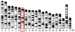
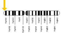
In human, the HBB gene is located on chromosome 11 at position p15.5.

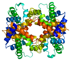
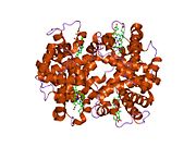
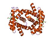
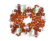
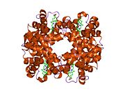
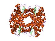
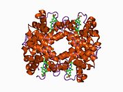
Hemoglobin subunit beta (beta globin, β-globin, haemoglobin beta, hemoglobin beta) is a globin protein, coded for by the HBB gene, which along with alpha globin (HBA), makes up the most common form of haemoglobin in adult humans, hemoglobin A (HbA). It is 147 amino acids long and has a molecular weight of 15,867 Da. Normal adult human HbA is a heterotetramer consisting of two alpha chains and two beta chains.

β-globin is encoded by the HBB gene on human chromosome 11. Mutations in the gene produce several variants of the proteins which are implicated with genetic disorders such as sickle-cell disease and beta thalassemia, as well as beneficial traits such as genetic resistance to malaria. At least 50 disease-causing mutations in this gene have been discovered.

Gene locus

Main article: Human β-globin locus

HBB protein is produced by the gene HBB which is located in the multigene locus of β-globin locus on chromosome 11, specifically on the short arm position 15.4. Expression of beta globin and the neighbouring globins in the β-globin locus is controlled by single locus control region (LCR), the most important regulatory element in the locus located upstream of the globin genes. The normal allelic variant is 1600 base pairs (bp) long and contains three exons. The order of the genes in the beta-globin cluster is 5' - epsilongamma-Ggamma-Adelta – beta - 3'.

Interactions

HBB interacts with Haemoglobin, alpha 1 (HBA1) to form haemoglobin A, the major haemoglobin in adult humans. The interaction is two-fold. First, one HBB and one HBA1 combine, non-covalently, to form a dimer. Secondly, two dimers combine to form the four-chain tetramer, and this becomes the functional haemoglobin.

Associated genetic disorders

Beta thalassemia

Beta thalassemia is an inherited genetic mutation in one (Beta thalassemia minor) or both (Beta thalassemia major) of the Beta globin alleles on chromosome 11. The mutant alleles are subdivided into two groups: β0, in which no functional β-globin is made, and β+, in which a small amount of normal β-globin protein is produced. Beta thalassemia minor occurs when an individual inherits one normal Beta allele and one abnormal Beta allele (either β0, or β+). Beta thalassemia minor results in a mild microcytic anemia that is often asymptomatic or may cause fatigue and or pale skin. Beta thalassemia major occurs when a person inherits two abnormal alleles. This can be either two β+ alleles, two β0 alleles, or one of each. Beta thalassemia major is a severe medical condition. A severe anemia is seen starting at 6 months of age. Without medical treatment death often occurs before age 12. Beta thalassemia major can be treated by lifelong blood transfusions or bone marrow transplantation.

According to a recent study, the stop gain mutation Gln40stop in HBB gene is a common cause of autosomal recessive Beta- thalassemia in Sardinian people (almost exclusive in Sardinia). Carriers of this mutation show an enhanced red blood cell count. As a curiosity, the same mutation was also associated to a decrease in serum LDL levels in carriers, so the authors suggest that is due to the need of cholesterol to regenerate cell membranes.

Sickle cell disease

More than a thousand naturally occurring HBB variants have been discovered. The most common is HbS, which causes sickle cell disease. HbS is produced by a point mutation in HBB in which the codon GAG is replaced by GTG. This results in the replacement of hydrophilic amino acid glutamic acid with the hydrophobic amino acid valine at the seventh position (β6Glu→Val). This substitution creates a hydrophobic spot on the outside of the protein that sticks to the hydrophobic region of an adjacent hemoglobin molecule's beta chain. This further causes clumping of HbS molecules into rigid fibers, causing "sickling" of the entire red blood cells in the homozygous (HbS/HbS) condition. The homozygous allele has become one of the deadliest genetic factors, whereas people heterozygous for the mutant allele (HbS/HbA) are resistant to malaria and develop minimal effects of the anaemia.

Haemoglobin C

Sickle cell disease is closely related to another mutant haemoglobin called haemoglobin C (HbC), because they can be inherited together. HbC mutation is at the same position in HbS, but glutamic acid is replaced by lysine (β6Glu→Lys). The mutation is particularly prevalent in West African populations. HbC provides near full protection against Plasmodium falciparum in homozygous (CC) individuals and intermediate protection in heterozygous (AC) individuals. This indicates that HbC has stronger influence than HbS, and is predicted to replace HbS in malaria-endemic regions.

Haemoglobin E

Another point mutation in HBB, in which glutamic acid is replaced with lysine at position 26 (β26Glu→Lys), leads to the formation of haemoglobin E (HbE). HbE has a very unstable α- and β-globin association. Even though the unstable protein itself has mild effect, inherited with HbS and thalassemia traits, it turns into a life-threatening form of β-thalassemia. The mutation is of relatively recent origin suggesting that it resulted from selective pressure against severe falciparum malaria, as heterozygous allele prevents the development of malaria.

Human evolution

Malaria due to Plasmodium falciparum is a major selective factor in human evolution. It has influenced mutations in HBB in various degrees resulting in the existence of numerous HBB variants. Some of these mutations are not directly lethal and instead confer resistance to malaria, particularly in Africa where malaria is epidemic. People of African descent have evolved to have higher rates of the mutant HBB because the heterozygous individuals have a misshaped red blood cell that prevent attacks from malarial parasites. Thus, HBB mutants are the sources of positive selection in these regions and are important for their long-term survival. Such selection markers are important for tracing human ancestry and diversification from Africa.

See also

References

  1. ^ GRCh38: Ensembl release 89: ENSG00000244734Ensembl, May 2017
  2. ^ GRCm38: Ensembl release 89: ENSMUSG00000073940Ensembl, May 2017
  3. "Human PubMed Reference:". National Center for Biotechnology Information, U.S. National Library of Medicine.
  4. "Mouse PubMed Reference:". National Center for Biotechnology Information, U.S. National Library of Medicine.
  5. ^ "Entrez Gene: HBB hemoglobin, beta".
  6. ^ Sabeti PC (2008). "Natural selection: uncovering mechanisms of evolutionary adaptation to infectious disease". Nature Education. 1 (1): 13.
  7. ^ Kwiatkowski DP (2005). "How malaria has affected the human genome and what human genetics can teach us about malaria". The American Journal of Human Genetics. 77 (2): 171–192. doi:10.1086/432519. PMC 1224522. PMID 16001361.
  8. Šimčíková D, Heneberg P (December 2019). "Refinement of evolutionary medicine predictions based on clinical evidence for the manifestations of Mendelian diseases". Scientific Reports. 9 (1): 18577. Bibcode:2019NatSR...918577S. doi:10.1038/s41598-019-54976-4. PMC 6901466. PMID 31819097.
  9. Levings PP, Bungert J (2002). "The human beta-globin locus control region". Eur. J. Biochem. 269 (6): 1589–99. doi:10.1046/j.1432-1327.2002.02797.x. PMID 11895428.
  10. Stelzl U, Worm U, Lalowski M, Haenig C, Brembeck FH, Goehler H, Stroedicke M, Zenkner M, Schoenherr A, Koeppen S, Timm J, Mintzlaff S, Abraham C, Bock N, Kietzmann S, Goedde A, Toksöz E, Droege A, Krobitsch S, Korn B, Birchmeier W, Lehrach H, Wanker EE (2005). "A human protein-protein interaction network: a resource for annotating the proteome". Cell. 122 (6): 957–968. doi:10.1016/j.cell.2005.08.029. hdl:11858/00-001M-0000-0010-8592-0. PMID 16169070. S2CID 8235923.
  11. Shaanan B (1983). "Structure of human oxyhaemoglobin at 2.1 A resolution". J. Mol. Biol. 171 (1). ENGLAND: 31–59. doi:10.1016/S0022-2836(83)80313-1. ISSN 0022-2836. PMID 6644819.
  12. "Hemoglobin Synthesis". harvard.edu. Harvard University. 2002. Retrieved 18 November 2014.
  13. H. Franklin Bunn, Vijay G. Sankaran (2017). "8". Pathology of blood disorders. pp. 927–933.
  14. Muncie HL, Campbell J (2009). "Alpha and beta thalassemia". American Family Physician. 80 (4): 339–44. PMID 19678601.
  15. "Beta thalassemia". Genetics Home Reference. U.S. National Library of Medicine. 11 November 2014. Retrieved 18 November 2014.
  16. Sidore, C., et al. (2015). "Genome sequencing elucidates Sardinian genetic architecture and augments association analyses for lipid and blood inflammatory markers". Nature Genetics. 47 (11): 1272–1281. doi:10.1038/ng.3368. PMC 4627508. PMID 26366554.
  17. Thom CS, Dickson CF, Gell DA, Weiss MJ (2013). "Hemoglobin variants: biochemical properties and clinical correlates". Cold Spring Harb Perspect Med. 3 (3): a011858. doi:10.1101/cshperspect.a011858. PMC 3579210. PMID 23388674.
  18. Lozano R, Naghavi M, Foreman K, Lim S, Shibuya K, Aboyans V, Abraham J, Adair T, Aggarwal R, Ahn SY, Alvarado M, Anderson HR, Anderson LM, Andrews KG, Atkinson C, Baddour LM, Barker-Collo S, Bartels DH, Bell ML, Benjamin EJ, Bennett D, Bhalla K, Bikbov B, Bin Abdulhak A, Birbeck G, Blyth F, Bolliger I, Boufous S, Bucello C, Burch M, Burney P, Carapetis J, Chen H, Chou D, Chugh SS, Coffeng LE, Colan SD, Colquhoun S, Colson KE, Condon J, Connor MD, Cooper LT, Corriere M, Cortinovis M, de Vaccaro KC, Couser W, Cowie BC, Criqui MH, Cross M, Dabhadkar KC, Dahodwala N, De Leo D, Degenhardt L, Delossantos A, Denenberg J, Des Jarlais DC, Dharmaratne SD, Dorsey ER, Driscoll T, Duber H, Ebel B, Erwin PJ, Espindola P, Ezzati M, Feigin V, Flaxman AD, Forouzanfar MH, Fowkes FG, Franklin R, Fransen M, Freeman MK, Gabriel SE, Gakidou E, Gaspari F, Gillum RF, Gonzalez-Medina D, Halasa YA, Haring D, Harrison JE, Havmoeller R, Hay RJ, Hoen B, Hotez PJ, Hoy D, Jacobsen KH, James SL, Jasrasaria R, Jayaraman S, Johns N, Karthikeyan G, Kassebaum N, Keren A, Khoo JP, Knowlton LM, Kobusingye O, Koranteng A, Krishnamurthi R, Lipnick M, Lipshultz SE, Ohno SL, Mabweijano J, MacIntyre MF, Mallinger L, March L, Marks GB, Marks R, Matsumori A, Matzopoulos R, Mayosi BM, McAnulty JH, McDermott MM, McGrath J, Mensah GA, Merriman TR, Michaud C, Miller M, Miller TR, Mock C, Mocumbi AO, Mokdad AA, Moran A, Mulholland K, Nair MN, Naldi L, Narayan KM, Nasseri K, Norman P, O'Donnell M, Omer SB, Ortblad K, Osborne R, Ozgediz D, Pahari B, Pandian JD, Rivero AP, Padilla RP, Perez-Ruiz F, Perico N, Phillips D, Pierce K, Pope CA, Porrini E, Pourmalek F, Raju M, Ranganathan D, Rehm JT, Rein DB, Remuzzi G, Rivara FP, Roberts T, De León FR, Rosenfeld LC, Rushton L, Sacco RL, Salomon JA, Sampson U, Sanman E, Schwebel DC, Segui-Gomez M, Shepard DS, Singh D, Singleton J, Sliwa K, Smith E, Steer A, Taylor JA, Thomas B, Tleyjeh IM, Towbin JA, Truelsen T, Undurraga EA, Venketasubramanian N, Vijayakumar L, Vos T, Wagner GR, Wang M, Wang W, Watt K, Weinstock MA, Weintraub R, Wilkinson JD, Woolf AD, Wulf S, Yeh PH, Yip P, Zabetian A, Zheng ZJ, Lopez AD, Murray CJ, AlMazroa MA, Memish ZA (2012). "Global and regional mortality from 235 causes of death for 20 age groups in 1990 and 2010: a systematic analysis for the Global Burden of Disease Study 2010". Lancet. 380 (9859): 2095–128. doi:10.1016/S0140-6736(12)61728-0. hdl:10536/DRO/DU:30050819. PMC 10790329. PMID 23245604. S2CID 1541253.
  19. Luzzatto L (2012). "Sickle cell anaemia and malaria". Mediterr J Hematol Infect Dis. 4 (1): e2012065. doi:10.4084/MJHID.2012.065. PMC 3499995. PMID 23170194.
  20. Piel FB, Howes RE, Patil AP, Nyangiri OA, Gething PW, Bhatt S, Williams TN, Weatherall DJ, Hay SI (2013). "The distribution of haemoglobin C and its prevalence in newborns in Africa". Scientific Reports. 3 (1671): 1671. Bibcode:2013NatSR...3E1671P. doi:10.1038/srep01671. PMC 3628164. PMID 23591685.
  21. Modiano D, Luoni G, Sirima BS, Simporé J, Verra F, Konaté A, Rastrelli E, Olivieri A, Calissano C, Paganotti GM, D'Urbano L, Sanou I, Sawadogo A, Modiano G, Coluzzi M (2001). "Haemoglobin C protects against clinical Plasmodium falciparum malaria". Nature. 414 (6861): 305–308. Bibcode:2001Natur.414..305M. doi:10.1038/35104556. PMID 11713529. S2CID 4360808.
  22. Verra F, Bancone G, Avellino P, Blot I, Simporé J, Modiano D (2007). "Haemoglobin C and S in natural selection against Plasmodium falciparum malaria: a plethora or a single shared adaptive mechanism?". Parassitologia. 49 (4): 209–13. PMID 18689228.
  23. Olivieri NF, Pakbaz Z, Vichinsky E (2011). "Hb E/beta-thalassaemia: a common & clinically diverse disorder". The Indian Journal of Medical Research. 134 (4): 522–531. PMC 3237252. PMID 22089616.
  24. Chotivanich K, Udomsangpetch R, Pattanapanyasat K, Chierakul W, Simpson J, Looareesuwan S, White N (2002). "Hemoglobin E: a balanced polymorphism protective against high parasitemias and thus severe P falciparum malaria". Blood. 100 (4): 1172–1176. doi:10.1182/blood.V100.4.1172.h81602001172_1172_1176. PMID 12149194.
  25. Verra F, Mangano VD, Modiano D (2009). "Genetics of susceptibility to Plasmodium falciparum: from classical malaria resistance genes towards genome-wide association studies". Parasite Immunology. 31 (5): 234–53. doi:10.1111/j.1365-3024.2009.01106.x. PMID 19388945. S2CID 23734166.
  26. Tishkoff SA, Williams SM (2002). "Genetic analysis of African populations: human evolution and complex disease". Nature Reviews Genetics. 3 (8): 611–21. doi:10.1038/nrg865. PMID 12154384. S2CID 7801737.
  27. Excoffier L (2002). "Human demographic history: refining the recent African origin model". Current Opinion in Genetics & Development. 12 (6): 675–682. doi:10.1016/S0959-437X(02)00350-7. PMID 12433581.
  28. Reed FA, Tishkoff SA (2006). "African human diversity, origins and migrations". Current Opinion in Genetics & Development. 16 (6): 597–605. doi:10.1016/j.gde.2006.10.008. PMID 17056248.

Further reading

External links

  • Overview of all the structural information available in the PDB for UniProt: P68871 (Human Hemoglobin subunit beta) at the PDBe-KB.
  • Overview of all the structural information available in the PDB for UniProt: P02088 (Mouse Hemoglobin subunit beta-1) at the PDBe-KB.
PDB gallery
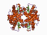
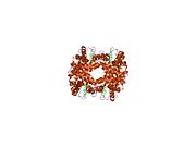
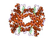
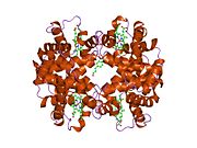
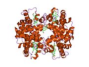
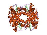
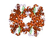
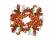
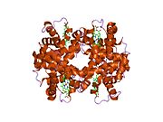
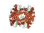
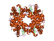
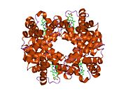
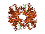
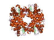
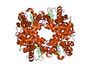
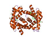
  • 1a00: HEMOGLOBIN (VAL BETA1 MET, TRP BETA37 TYR) MUTANT 1a00: HEMOGLOBIN (VAL BETA1 MET, TRP BETA37 TYR) MUTANT
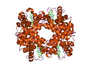
  • 1a01: HEMOGLOBIN (VAL BETA1 MET, TRP BETA37 ALA) MUTANT 1a01: HEMOGLOBIN (VAL BETA1 MET, TRP BETA37 ALA) MUTANT
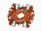
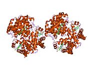
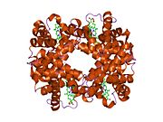
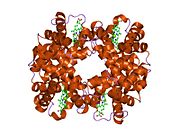
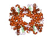
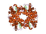
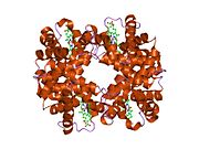
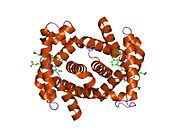
  • 1a0u: HEMOGLOBIN (VAL BETA1 MET) MUTANT 1a0u: HEMOGLOBIN (VAL BETA1 MET) MUTANT
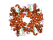
  • 1a0z: HEMOGLOBIN (VAL BETA1 MET) MUTANT 1a0z: HEMOGLOBIN (VAL BETA1 MET) MUTANT
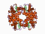
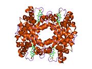
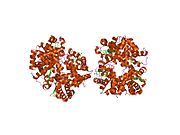
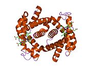
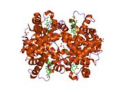
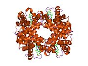
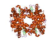
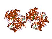
  • 1a3n: DEOXY HUMAN HEMOGLOBIN 1a3n: DEOXY HUMAN HEMOGLOBIN
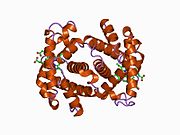
  • 1a3o: ARTIFICIAL MUTANT (ALPHA Y42H) OF DEOXY HEMOGLOBIN 1a3o: ARTIFICIAL MUTANT (ALPHA Y42H) OF DEOXY HEMOGLOBIN
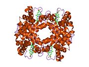
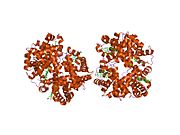
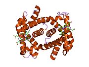
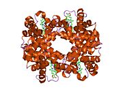
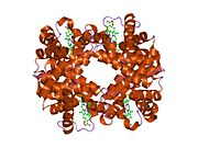
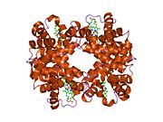
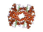
  • 1abw: DEOXY RHB1.1 (RECOMBINANT HEMOGLOBIN) 1abw: DEOXY RHB1.1 (RECOMBINANT HEMOGLOBIN)
  • 1aby: CYANOMET RHB1.1 (RECOMBINANT HEMOGLOBIN) 1aby: CYANOMET RHB1.1 (RECOMBINANT HEMOGLOBIN)
  • 1aj9: R-STATE HUMAN CARBONMONOXYHEMOGLOBIN ALPHA-A53S 1aj9: R-STATE HUMAN CARBONMONOXYHEMOGLOBIN ALPHA-A53S
  • 1b86: HUMAN DEOXYHAEMOGLOBIN-2,3-DIPHOSPHOGLYCERATE COMPLEX 1b86: HUMAN DEOXYHAEMOGLOBIN-2,3-DIPHOSPHOGLYCERATE COMPLEX
  • 1bab: HEMOGLOBIN THIONVILLE: AN ALPHA-CHAIN VARIANT WITH A SUBSTITUTION OF A GLUTAMATE FOR VALINE AT NA-1 AND HAVING AN ACETYLATED METHIONINE NH2 TERMINUS 1bab: HEMOGLOBIN THIONVILLE: AN ALPHA-CHAIN VARIANT WITH A SUBSTITUTION OF A GLUTAMATE FOR VALINE AT NA-1 AND HAVING AN ACETYLATED METHIONINE NH2 TERMINUS
  • 1bbb: A THIRD QUATERNARY STRUCTURE OF HUMAN HEMOGLOBIN A AT 1.7-ANGSTROMS RESOLUTION 1bbb: A THIRD QUATERNARY STRUCTURE OF HUMAN HEMOGLOBIN A AT 1.7-ANGSTROMS RESOLUTION
  • 1bij: CROSSLINKED, DEOXY HUMAN HEMOGLOBIN A 1bij: CROSSLINKED, DEOXY HUMAN HEMOGLOBIN A
  • 1buw: CRYSTAL STRUCTURE OF S-NITROSO-NITROSYL HUMAN HEMOGLOBIN A 1buw: CRYSTAL STRUCTURE OF S-NITROSO-NITROSYL HUMAN HEMOGLOBIN A
  • 1bz0: HEMOGLOBIN A (HUMAN, DEOXY, HIGH SALT) 1bz0: HEMOGLOBIN A (HUMAN, DEOXY, HIGH SALT)
  • 1bz1: HEMOGLOBIN (ALPHA + MET) VARIANT 1bz1: HEMOGLOBIN (ALPHA + MET) VARIANT
  • 1bzz: HEMOGLOBIN (ALPHA V1M) MUTANT 1bzz: HEMOGLOBIN (ALPHA V1M) MUTANT
  • 1c7b: DEOXY RHB1.0 (RECOMBINANT HEMOGLOBIN) 1c7b: DEOXY RHB1.0 (RECOMBINANT HEMOGLOBIN)
  • 1c7c: DEOXY RHB1.1 (RECOMBINANT HEMOGLOBIN) 1c7c: DEOXY RHB1.1 (RECOMBINANT HEMOGLOBIN)
  • 1c7d: DEOXY RHB1.2 (RECOMBINANT HEMOGLOBIN) 1c7d: DEOXY RHB1.2 (RECOMBINANT HEMOGLOBIN)
  • 1cbl: THE 1.9 ANGSTROM STRUCTURE OF DEOXY-BETA4 HEMOGLOBIN: ANALYSIS OF THE PARTITIONING OF QUATERNARY-ASSOCIATED AND LIGAND-INDUCED CHANGES IN TERTIARY STRUCTURE 1cbl: THE 1.9 ANGSTROM STRUCTURE OF DEOXY-BETA4 HEMOGLOBIN: ANALYSIS OF THE PARTITIONING OF QUATERNARY-ASSOCIATED AND LIGAND-INDUCED CHANGES IN TERTIARY STRUCTURE
  • 1cbm: THE 1.8 ANGSTROM STRUCTURE OF CARBONMONOXY-BETA4 HEMOGLOBIN: ANALYSIS OF A HOMOTETRAMER WITH THE R QUATERNARY STRUCTURE OF LIGANDED ALPHA2BETA2 HEMOGLOBIN 1cbm: THE 1.8 ANGSTROM STRUCTURE OF CARBONMONOXY-BETA4 HEMOGLOBIN: ANALYSIS OF A HOMOTETRAMER WITH THE R QUATERNARY STRUCTURE OF LIGANDED ALPHA2BETA2 HEMOGLOBIN
  • 1cls: CROSS-LINKED HUMAN HEMOGLOBIN DEOXY 1cls: CROSS-LINKED HUMAN HEMOGLOBIN DEOXY
  • 1cmy: THE MUTATION BETA99 ASP-TYR STABILIZES Y-A NEW, COMPOSITE QUATERNARY STATE OF HUMAN HEMOGLOBIN 1cmy: THE MUTATION BETA99 ASP-TYR STABILIZES Y-A NEW, COMPOSITE QUATERNARY STATE OF HUMAN HEMOGLOBIN
  • 1coh: STRUCTURE OF HAEMOGLOBIN IN THE DEOXY QUATERNARY STATE WITH LIGAND BOUND AT THE ALPHA HAEMS 1coh: STRUCTURE OF HAEMOGLOBIN IN THE DEOXY QUATERNARY STATE WITH LIGAND BOUND AT THE ALPHA HAEMS
  • 1dke: NI BETA HEME HUMAN HEMOGLOBIN 1dke: NI BETA HEME HUMAN HEMOGLOBIN
  • 1dxt: HIGH-RESOLUTION X-RAY STUDY OF DEOXY RECOMBINANT HUMAN HEMOGLOBINS SYNTHESIZED FROM BETA-GLOBINS HAVING MUTATED AMINO TERMINI 1dxt: HIGH-RESOLUTION X-RAY STUDY OF DEOXY RECOMBINANT HUMAN HEMOGLOBINS SYNTHESIZED FROM BETA-GLOBINS HAVING MUTATED AMINO TERMINI
  • 1dxu: HIGH-RESOLUTION X-RAY STUDY OF DEOXY RECOMBINANT HUMAN HEMOGLOBINS SYNTHESIZED FROM BETA-GLOBINS HAVING MUTATED AMINO TERMINI 1dxu: HIGH-RESOLUTION X-RAY STUDY OF DEOXY RECOMBINANT HUMAN HEMOGLOBINS SYNTHESIZED FROM BETA-GLOBINS HAVING MUTATED AMINO TERMINI
  • 1dxv: HIGH-RESOLUTION X-RAY STUDY OF DEOXY RECOMBINANT HUMAN HEMOGLOBINS SYNTHESIZED FROM BETA-GLOBINS HAVING MUTATED AMINO TERMINI 1dxv: HIGH-RESOLUTION X-RAY STUDY OF DEOXY RECOMBINANT HUMAN HEMOGLOBINS SYNTHESIZED FROM BETA-GLOBINS HAVING MUTATED AMINO TERMINI
  • 1fn3: CRYSTAL STRUCTURE OF NICKEL RECONSTITUTED HEMOGLOBIN-A CASE FOR PERMANENT, T-STATE HEMOGLOBIN 1fn3: CRYSTAL STRUCTURE OF NICKEL RECONSTITUTED HEMOGLOBIN-A CASE FOR PERMANENT, T-STATE HEMOGLOBIN
  • 1g9v: HIGH RESOLUTION CRYSTAL STRUCTURE OF DEOXY HEMOGLOBIN COMPLEXED WITH A POTENT ALLOSTERIC EFFECTOR 1g9v: HIGH RESOLUTION CRYSTAL STRUCTURE OF DEOXY HEMOGLOBIN COMPLEXED WITH A POTENT ALLOSTERIC EFFECTOR
  • 1gbu: DEOXY (BETA-(C93A,C112G)) HUMAN HEMOGLOBIN 1gbu: DEOXY (BETA-(C93A,C112G)) HUMAN HEMOGLOBIN
  • 1gbv: (ALPHA-OXY, BETA-(C112G)DEOXY) T-STATE HUMAN HEMOGLOBIN 1gbv: (ALPHA-OXY, BETA-(C112G)DEOXY) T-STATE HUMAN HEMOGLOBIN
  • 1gli: DEOXYHEMOGLOBIN T38W (ALPHA CHAINS), V1G (ALPHA AND BETA CHAINS) 1gli: DEOXYHEMOGLOBIN T38W (ALPHA CHAINS), V1G (ALPHA AND BETA CHAINS)
  • 1gzx: OXY T STATE HAEMOGLOBIN: OXYGEN BOUND AT ALL FOUR HAEMS 1gzx: OXY T STATE HAEMOGLOBIN: OXYGEN BOUND AT ALL FOUR HAEMS
  • 1hab: CROSSLINKED HAEMOGLOBIN 1hab: CROSSLINKED HAEMOGLOBIN
  • 1hac: CROSSLINKED HAEMOGLOBIN 1hac: CROSSLINKED HAEMOGLOBIN
  • 1hba: HIGH-RESOLUTION X-RAY STUDY OF DEOXYHEMOGLOBIN ROTHSCHILD 37BETA TRP-> ARG: A MUTATION THAT CREATES AN INTERSUBUNIT CHLORIDE-BINDING SITE 1hba: HIGH-RESOLUTION X-RAY STUDY OF DEOXYHEMOGLOBIN ROTHSCHILD 37BETA TRP-> ARG: A MUTATION THAT CREATES AN INTERSUBUNIT CHLORIDE-BINDING SITE
  • 1hbb: HIGH-RESOLUTION X-RAY STUDY OF DEOXYHEMOGLOBIN ROTHSCHILD 37BETA TRP-> ARG: A MUTATION THAT CREATES AN INTERSUBUNIT CHLORIDE-BINDING SITE 1hbb: HIGH-RESOLUTION X-RAY STUDY OF DEOXYHEMOGLOBIN ROTHSCHILD 37BETA TRP-> ARG: A MUTATION THAT CREATES AN INTERSUBUNIT CHLORIDE-BINDING SITE
  • 1hbs: REFINED CRYSTAL STRUCTURE OF DEOXYHEMOGLOBIN S. I. RESTRAINED LEAST-SQUARES REFINEMENT AT 3.0-ANGSTROMS RESOLUTION 1hbs: REFINED CRYSTAL STRUCTURE OF DEOXYHEMOGLOBIN S. I. RESTRAINED LEAST-SQUARES REFINEMENT AT 3.0-ANGSTROMS RESOLUTION
  • 1hco: THE STRUCTURE OF HUMAN CARBONMONOXY HAEMOGLOBIN AT 2.7 ANGSTROMS RESOLUTION 1hco: THE STRUCTURE OF HUMAN CARBONMONOXY HAEMOGLOBIN AT 2.7 ANGSTROMS RESOLUTION
  • 1hdb: ANALYSIS OF THE CRYSTAL STRUCTURE, MOLECULAR MODELING AND INFRARED SPECTROSCOPY OF THE DISTAL BETA-HEME POCKET VALINE67(E11)-THREONINE MUTATION OF HEMOGLOBIN 1hdb: ANALYSIS OF THE CRYSTAL STRUCTURE, MOLECULAR MODELING AND INFRARED SPECTROSCOPY OF THE DISTAL BETA-HEME POCKET VALINE67(E11)-THREONINE MUTATION OF HEMOGLOBIN
  • 1hga: HIGH RESOLUTION CRYSTAL STRUCTURES AND COMPARISONS OF T STATE DEOXYHAEMOGLOBIN AND TWO LIGANDED T-STATE HAEMOGLOBINS: T(ALPHA-OXY)HAEMOGLOBIN AND T(MET)HAEMOGLOBIN 1hga: HIGH RESOLUTION CRYSTAL STRUCTURES AND COMPARISONS OF T STATE DEOXYHAEMOGLOBIN AND TWO LIGANDED T-STATE HAEMOGLOBINS: T(ALPHA-OXY)HAEMOGLOBIN AND T(MET)HAEMOGLOBIN
  • 1hgb: HIGH RESOLUTION CRYSTAL STRUCTURES AND COMPARISONS OF T STATE DEOXYHAEMOGLOBIN AND TWO LIGANDED T-STATE HAEMOGLOBINS: T(ALPHA-OXY)HAEMOGLOBIN AND T(MET)HAEMOGLOBIN 1hgb: HIGH RESOLUTION CRYSTAL STRUCTURES AND COMPARISONS OF T STATE DEOXYHAEMOGLOBIN AND TWO LIGANDED T-STATE HAEMOGLOBINS: T(ALPHA-OXY)HAEMOGLOBIN AND T(MET)HAEMOGLOBIN
  • 1hgc: HIGH RESOLUTION CRYSTAL STRUCTURES AND COMPARISONS OF T STATE DEOXYHAEMOGLOBIN AND TWO LIGANDED T-STATE HAEMOGLOBINS: T(ALPHA-OXY)HAEMOGLOBIN AND T(MET)HAEMOGLOBIN 1hgc: HIGH RESOLUTION CRYSTAL STRUCTURES AND COMPARISONS OF T STATE DEOXYHAEMOGLOBIN AND TWO LIGANDED T-STATE HAEMOGLOBINS: T(ALPHA-OXY)HAEMOGLOBIN AND T(MET)HAEMOGLOBIN
  • 1hho: STRUCTURE OF HUMAN OXYHAEMOGLOBIN AT 2.1 ANGSTROMS RESOLUTION 1hho: STRUCTURE OF HUMAN OXYHAEMOGLOBIN AT 2.1 ANGSTROMS RESOLUTION
  • 1ird: Crystal Structure of Human Carbonmonoxy-Haemoglobin at 1.25 A Resolution 1ird: Crystal Structure of Human Carbonmonoxy-Haemoglobin at 1.25 A Resolution
  • 1j3y: Direct observation of photolysis-induced tertiary structural changes in human hemoglobin; Crystal structure of alpha(Fe)-beta(Ni) hemoglobin (laser photolysed) 1j3y: Direct observation of photolysis-induced tertiary structural changes in human hemoglobin; Crystal structure of alpha(Fe)-beta(Ni) hemoglobin (laser photolysed)
  • 1j3z: Direct observation of photolysis-induced tertiary structural changes in human haemoglobin; Crystal structure of alpha(Fe-CO)-beta(Ni) hemoglobin (laser unphotolysed) 1j3z: Direct observation of photolysis-induced tertiary structural changes in human haemoglobin; Crystal structure of alpha(Fe-CO)-beta(Ni) hemoglobin (laser unphotolysed)
  • 1j40: Direct observation of photolysis-induced tertiary structural changes in human haemoglobin; Crystal structure of alpha(Ni)-beta(Fe-CO) hemoglobin (laser unphotolysed) 1j40: Direct observation of photolysis-induced tertiary structural changes in human haemoglobin; Crystal structure of alpha(Ni)-beta(Fe-CO) hemoglobin (laser unphotolysed)
  • 1j41: Direct observation of photolysis-induced tertiary structural changes in human haemoglobin; Crystal structure of alpha(Ni)-beta(Fe) hemoglobin (laser photolysed) 1j41: Direct observation of photolysis-induced tertiary structural changes in human haemoglobin; Crystal structure of alpha(Ni)-beta(Fe) hemoglobin (laser photolysed)
  • 1j7s: Crystal Structure of deoxy HbalphaYQ, a mutant of HbA 1j7s: Crystal Structure of deoxy HbalphaYQ, a mutant of HbA
  • 1j7w: Crystal structure of deoxy HbbetaYQ, a site directed mutant of HbA 1j7w: Crystal structure of deoxy HbbetaYQ, a site directed mutant of HbA
  • 1j7y: Crystal structure of partially ligated mutant of HbA 1j7y: Crystal structure of partially ligated mutant of HbA
  • 1jy7: THE STRUCTURE OF HUMAN METHEMOGLOBIN. THE VARIATION OF A THEME 1jy7: THE STRUCTURE OF HUMAN METHEMOGLOBIN. THE VARIATION OF A THEME
  • 1k0y: X-ray Crystallographic Analyses of Symmetrical Allosteric Effectors of Hemoglobin. Compounds Designed to Link Primary and Secondary Binding Sites 1k0y: X-ray Crystallographic Analyses of Symmetrical Allosteric Effectors of Hemoglobin. Compounds Designed to Link Primary and Secondary Binding Sites
  • 1k1k: Structure of Mutant Human Carbonmonoxyhemoglobin C (beta E6K) at 2.0 Angstrom Resolution in Phosphate Buffer. 1k1k: Structure of Mutant Human Carbonmonoxyhemoglobin C (beta E6K) at 2.0 Angstrom Resolution in Phosphate Buffer.
  • 1kd2: Crystal Structure of Human Deoxyhemoglobin in Absence of Any Anions 1kd2: Crystal Structure of Human Deoxyhemoglobin in Absence of Any Anions
  • 1lfl: DEOXY HEMOGLOBIN (90% RELATIVE HUMIDITY) 1lfl: DEOXY HEMOGLOBIN (90% RELATIVE HUMIDITY)
  • 1lfq: OXY HEMOGLOBIN (93% RELATIVE HUMIDITY) 1lfq: OXY HEMOGLOBIN (93% RELATIVE HUMIDITY)
  • 1lft: OXY HEMOGLOBIN (90% RELATIVE HUMIDITY) 1lft: OXY HEMOGLOBIN (90% RELATIVE HUMIDITY)
  • 1lfv: OXY HEMOGLOBIN (88% RELATIVE HUMIDITY) 1lfv: OXY HEMOGLOBIN (88% RELATIVE HUMIDITY)
  • 1lfy: OXY HEMOGLOBIN (84% RELATIVE HUMIDITY) 1lfy: OXY HEMOGLOBIN (84% RELATIVE HUMIDITY)
  • 1lfz: OXY HEMOGLOBIN (25% METHANOL) 1lfz: OXY HEMOGLOBIN (25% METHANOL)
  • 1ljw: Crystal Structure of Human Carbonmonoxy Hemoglobin at 2.16 A: A Snapshot of the Allosteric Transition 1ljw: Crystal Structure of Human Carbonmonoxy Hemoglobin at 2.16 A: A Snapshot of the Allosteric Transition
  • 1m9p: Crystalline Human Carbonmonoxy Hemoglobin C Exhibits The R2 Quaternary State at Neutral pH In The Presence of Polyethylene Glycol: The 2.1 Angstrom Resolution Crystal Structure 1m9p: Crystalline Human Carbonmonoxy Hemoglobin C Exhibits The R2 Quaternary State at Neutral pH In The Presence of Polyethylene Glycol: The 2.1 Angstrom Resolution Crystal Structure
  • 1mko: A Fourth Quaternary Structure of Human Hemoglobin A at 2.18 A Resolution 1mko: A Fourth Quaternary Structure of Human Hemoglobin A at 2.18 A Resolution
  • 1nej: Crystalline Human Carbonmonoxy Hemoglobin S (Liganded Sickle Cell Hemoglobin) Exhibits The R2 Quaternary State At Neutral pH In The Presence Of Polyethylene Glycol: The 2.1 Angstrom Resolution Crystal Structure 1nej: Crystalline Human Carbonmonoxy Hemoglobin S (Liganded Sickle Cell Hemoglobin) Exhibits The R2 Quaternary State At Neutral pH In The Presence Of Polyethylene Glycol: The 2.1 Angstrom Resolution Crystal Structure
  • 1nih: Structure of deoxy-quaternary haemoglobin with liganded beta subunits 1nih: Structure of deoxy-quaternary haemoglobin with liganded beta subunits
  • 1nqp: Crystal structure of Human hemoglobin E at 1.73 A resolution 1nqp: Crystal structure of Human hemoglobin E at 1.73 A resolution
  • 1o1i: Cyanomet hemoglobin (A-GLY-C:V1M,L29F,H58Q; B,D:V1M,L106W) 1o1i: Cyanomet hemoglobin (A-GLY-C:V1M,L29F,H58Q; B,D:V1M,L106W)
  • 1o1j: Deoxy hemoglobin (A-GLY-C:V1M,L29F,H58Q; B,D:V1M,L106W) 1o1j: Deoxy hemoglobin (A-GLY-C:V1M,L29F,H58Q; B,D:V1M,L106W)
  • 1o1k: Deoxy hemoglobin (A,C:V1M; B,D:V1M,V67W) 1o1k: Deoxy hemoglobin (A,C:V1M; B,D:V1M,V67W)
  • 1o1l: Deoxy hemoglobin (A-GLY-C:V1M,L29W,H58Q; B,D:V1M) 1o1l: Deoxy hemoglobin (A-GLY-C:V1M,L29W,H58Q; B,D:V1M)
  • 1o1m: Deoxy hemoglobin (A-GLYGLYGLY-C:V1M,L29F,H58Q B,D:V1M,V67W) 1o1m: Deoxy hemoglobin (A-GLYGLYGLY-C:V1M,L29F,H58Q B,D:V1M,V67W)
  • 1o1n: Deoxy hemoglobin (A-GLYGLYGLY-C:V1M,L29W; B,D:V1M) 1o1n: Deoxy hemoglobin (A-GLYGLYGLY-C:V1M,L29W; B,D:V1M)
  • 1o1o: Deoxy hemoglobin (A,C:V1M,V62L; B,D:V1M,V67L) 1o1o: Deoxy hemoglobin (A,C:V1M,V62L; B,D:V1M,V67L)
  • 1o1p: Deoxy hemoglobin (A-GLY-C:V1M; B,D:V1M,C93A,N108K) 1o1p: Deoxy hemoglobin (A-GLY-C:V1M; B,D:V1M,C93A,N108K)
  • 1qi8: DEOXYGENATED STRUCTURE OF A DISTAL POCKET HEMOGLOBIN MUTANT 1qi8: DEOXYGENATED STRUCTURE OF A DISTAL POCKET HEMOGLOBIN MUTANT
  • 1qsh: MAGNESIUM(II)-AND ZINC(II)-PROTOPORPHYRIN IX'S STABILIZE THE LOWEST OXYGEN AFFINITY STATE OF HUMAN HEMOGLOBIN EVEN MORE STRONGLY THAN DEOXYHEME 1qsh: MAGNESIUM(II)-AND ZINC(II)-PROTOPORPHYRIN IX'S STABILIZE THE LOWEST OXYGEN AFFINITY STATE OF HUMAN HEMOGLOBIN EVEN MORE STRONGLY THAN DEOXYHEME
  • 1qsi: MAGNESIUM(II)-AND ZINC(II)-PROTOPORPHYRIN IX'S STABILIZE THE LOWEST OXYGEN AFFINITY STATE OF HUMAN HEMOGLOBIN EVEN MORE STRONGLY THAN DEOXYHEME 1qsi: MAGNESIUM(II)-AND ZINC(II)-PROTOPORPHYRIN IX'S STABILIZE THE LOWEST OXYGEN AFFINITY STATE OF HUMAN HEMOGLOBIN EVEN MORE STRONGLY THAN DEOXYHEME
  • 1qxd: Structural Basis for the Potent Antisickling Effect of a Novel Class of 5-Membered Heterocyclic Aldehydic Compounds 1qxd: Structural Basis for the Potent Antisickling Effect of a Novel Class of 5-Membered Heterocyclic Aldehydic Compounds
  • 1qxe: Structural Basis for the Potent Antisickling Effect of a Novel Class of 5-Membered Heterocyclic Aldehydic Compounds 1qxe: Structural Basis for the Potent Antisickling Effect of a Novel Class of 5-Membered Heterocyclic Aldehydic Compounds
  • 1r1x: Crystal structure of oxy-human hemoglobin Bassett at 2.15 angstrom 1r1x: Crystal structure of oxy-human hemoglobin Bassett at 2.15 angstrom
  • 1r1y: Crystal structure of deoxy-human hemoglobin Bassett at 1.8 angstrom 1r1y: Crystal structure of deoxy-human hemoglobin Bassett at 1.8 angstrom
  • 1rps: Crystallographic Analysis of the Interaction of Nitric Oxide with Quaternary-T Human Hemoglobin. Hemoglobin exposed to NO under anerobic conditions 1rps: Crystallographic Analysis of the Interaction of Nitric Oxide with Quaternary-T Human Hemoglobin. Hemoglobin exposed to NO under anerobic conditions
  • 1rq3: Crystallographic Analysis of the Interaction of Nitric Oxide with Quaternary-T Human Deoxyhemoglobin, Deoxyhemoglobin 1rq3: Crystallographic Analysis of the Interaction of Nitric Oxide with Quaternary-T Human Deoxyhemoglobin, Deoxyhemoglobin
  • 1rq4: Crystallographic Analysis of the Interaction of Nitric Oxide with Quaternary-T Human Hemoglobin, HEMOGLOBIN EXPOSED TO NO UNDER AEROBIC CONDITIONS 1rq4: Crystallographic Analysis of the Interaction of Nitric Oxide with Quaternary-T Human Hemoglobin, HEMOGLOBIN EXPOSED TO NO UNDER AEROBIC CONDITIONS
  • 1rqa: Crystallographic Analysis of the Interaction of Nitric Oxide with Quaternary-T Human Hemoglobin. Beta W73E hemoglobin exposed to NO under anaerobic conditions 1rqa: Crystallographic Analysis of the Interaction of Nitric Oxide with Quaternary-T Human Hemoglobin. Beta W73E hemoglobin exposed to NO under anaerobic conditions
  • 1rvw: R STATE HUMAN HEMOGLOBIN , CARBONMONOXY 1rvw: R STATE HUMAN HEMOGLOBIN , CARBONMONOXY
  • 1sdk: CROSS-LINKED, CARBONMONOXY HEMOGLOBIN A 1sdk: CROSS-LINKED, CARBONMONOXY HEMOGLOBIN A
  • 1sdl: CROSS-LINKED, CARBONMONOXY HEMOGLOBIN A 1sdl: CROSS-LINKED, CARBONMONOXY HEMOGLOBIN A
  • 1shr: Crystal structure of ferrocyanide bound human hemoglobin A2 at 1.88A resolution 1shr: Crystal structure of ferrocyanide bound human hemoglobin A2 at 1.88A resolution
  • 1si4: Crystal structure of Human hemoglobin A2 (in R2 state) at 2.2 A resolution 1si4: Crystal structure of Human hemoglobin A2 (in R2 state) at 2.2 A resolution
  • 1thb: REFINEMENT OF A PARTIALLY OXYGENATED T STATE HAEMOGLOBIN AT 1.5 ANGSTROMS RESOLUTION 1thb: REFINEMENT OF A PARTIALLY OXYGENATED T STATE HAEMOGLOBIN AT 1.5 ANGSTROMS RESOLUTION
  • 1uiw: Crystal Structures of Unliganded and Half-Liganded Human Hemoglobin Derivatives Cross-Linked between Lys 82beta1 and Lys 82beta2 1uiw: Crystal Structures of Unliganded and Half-Liganded Human Hemoglobin Derivatives Cross-Linked between Lys 82beta1 and Lys 82beta2
  • 1vwt: T STATE HUMAN HEMOGLOBIN , ALPHA AQUOMET, BETA DEOXY 1vwt: T STATE HUMAN HEMOGLOBIN , ALPHA AQUOMET, BETA DEOXY
  • 1xxt: The T-to-T High Transitions in Human Hemoglobin: wild-type deoxy Hb A (low salt, one test set) 1xxt: The T-to-T High Transitions in Human Hemoglobin: wild-type deoxy Hb A (low salt, one test set)
  • 1xy0: T-to-THigh Transitions in Human Hemoglobin: alphaK40G deoxy low-salt 1xy0: T-to-THigh Transitions in Human Hemoglobin: alphaK40G deoxy low-salt
  • 1xye: T-to-THigh Transitions in Human Hemoglobin: alpha Y42A deoxy low salt 1xye: T-to-THigh Transitions in Human Hemoglobin: alpha Y42A deoxy low salt
  • 1xz2: wild-type hemoglobin deoxy no-salt 1xz2: wild-type hemoglobin deoxy no-salt
  • 1xz4: Intersubunit Interactions Associated with Tyr42alpha Stabilize the Quaternary-T Tetramer but are not Major Quaternary Constraints in Deoxyhemoglobin: alphaY42A deoxyhemoglobin no-salt 1xz4: Intersubunit Interactions Associated with Tyr42alpha Stabilize the Quaternary-T Tetramer but are not Major Quaternary Constraints in Deoxyhemoglobin: alphaY42A deoxyhemoglobin no-salt
  • 1xz5: T-to-THigh Quaternary Transitions in Human Hemoglobin: alphaL91A deoxy low-salt 1xz5: T-to-THigh Quaternary Transitions in Human Hemoglobin: alphaL91A deoxy low-salt
  • 1xz7: T-to-THigh Quaternary Transitions in Human Hemoglobin: alphaR92A deoxy low-salt 1xz7: T-to-THigh Quaternary Transitions in Human Hemoglobin: alphaR92A deoxy low-salt
  • 1xzu: T-to-THigh Quaternary Transitions in Human Hemoglobin: alphaD94G deoxy low-salt 1xzu: T-to-THigh Quaternary Transitions in Human Hemoglobin: alphaD94G deoxy low-salt
  • 1xzv: T-to-THigh Quaternary Transitions in Human Hemoglobin: alphaP95A deoxy low-salt 1xzv: T-to-THigh Quaternary Transitions in Human Hemoglobin: alphaP95A deoxy low-salt
  • 1y09: T-to-T(High) Quaternary Transitions in Human Hemoglobin: alphaN97A deoxy low-salt 1y09: T-to-T(High) Quaternary Transitions in Human Hemoglobin: alphaN97A deoxy low-salt
  • 1y0a: T-to-THigh Quaternary Transitions in Human Hemoglobin: alphaY140A deoxy low-salt 1y0a: T-to-THigh Quaternary Transitions in Human Hemoglobin: alphaY140A deoxy low-salt
  • 1y0c: T-to-THigh Quaternary Transitions in Human Hemoglobin: alphaY140F deoxy low-salt 1y0c: T-to-THigh Quaternary Transitions in Human Hemoglobin: alphaY140F deoxy low-salt
  • 1y0d: T-to-THigh Quaternary Transitions in Human Hemoglobin: desArg141alpha deoxy low-salt 1y0d: T-to-THigh Quaternary Transitions in Human Hemoglobin: desArg141alpha deoxy low-salt
  • 1y0t: T-to-T(High) Quaternary Transitions in Human Hemoglobin: betaV1M deoxy low-salt (1 test set) 1y0t: T-to-T(High) Quaternary Transitions in Human Hemoglobin: betaV1M deoxy low-salt (1 test set)
  • 1y0w: T-to-THigh quaternary Transitions in Human Hemoglobin: betaV1M deoxy low-salt (10 test sets) 1y0w: T-to-THigh quaternary Transitions in Human Hemoglobin: betaV1M deoxy low-salt (10 test sets)
  • 1y22: T-To-T(High) quaternary transitions in human hemoglobin: betaV33A deoxy low-salt (1 test set) 1y22: T-To-T(High) quaternary transitions in human hemoglobin: betaV33A deoxy low-salt (1 test set)
  • 1y2z: T-To-T(High) quaternary transitions in human hemoglobin: betaV34G deoxy low-salt (1 test set) 1y2z: T-To-T(High) quaternary transitions in human hemoglobin: betaV34G deoxy low-salt (1 test set)
  • 1y31: T-To-T(High) quaternary transitions in human hemoglobin: betaY35A deoxy low-salt (1 test set) 1y31: T-To-T(High) quaternary transitions in human hemoglobin: betaY35A deoxy low-salt (1 test set)
  • 1y35: T-To-T(High) quaternary transitions in human hemoglobin: betaY35F deoxy low-salt (1 test set) 1y35: T-To-T(High) quaternary transitions in human hemoglobin: betaY35F deoxy low-salt (1 test set)
  • 1y45: T-To-T(high) quaternary transitions in human hemoglobin: betaP36A deoxy low-salt (10 test sets) 1y45: T-To-T(high) quaternary transitions in human hemoglobin: betaP36A deoxy low-salt (10 test sets)
  • 1y46: T-To-T(High) quaternary transitions in human hemoglobin: betaW37Y deoxy low-salt (10 test sets) 1y46: T-To-T(High) quaternary transitions in human hemoglobin: betaW37Y deoxy low-salt (10 test sets)
  • 1y4b: T-To-T(High) quaternary transitions in human hemoglobin: betaW37H deoxy low-salt (10 test sets) 1y4b: T-To-T(High) quaternary transitions in human hemoglobin: betaW37H deoxy low-salt (10 test sets)
  • 1y4f: T-To-T(High) quaternary transitions in human hemoglobin: betaW37A deoxy low-salt (10 test sets) 1y4f: T-To-T(High) quaternary transitions in human hemoglobin: betaW37A deoxy low-salt (10 test sets)
  • 1y4g: T-To-T(High) quaternary transitions in human hemoglobin: betaW37G deoxy low-salt (10 test sets) 1y4g: T-To-T(High) quaternary transitions in human hemoglobin: betaW37G deoxy low-salt (10 test sets)
  • 1y4p: T-To-T(high) quaternary transitions in human hemoglobin: betaW37E deoxy low-salt (10 test sets) 1y4p: T-To-T(high) quaternary transitions in human hemoglobin: betaW37E deoxy low-salt (10 test sets)
  • 1y4q: T-To-T(High) quaternary transitions in human hemoglobin: betaF42A deoxy low-salt (1 test set) 1y4q: T-To-T(High) quaternary transitions in human hemoglobin: betaF42A deoxy low-salt (1 test set)
  • 1y4r: T-To-T(High) quaternary transitions in human hemoglobin: betaF45A deoxy low-salt (1 test set) 1y4r: T-To-T(High) quaternary transitions in human hemoglobin: betaF45A deoxy low-salt (1 test set)
  • 1y4v: T-To-T(High) quaternary transitions in human hemoglobin: betaC93A deoxy low-salt (1 test set) 1y4v: T-To-T(High) quaternary transitions in human hemoglobin: betaC93A deoxy low-salt (1 test set)
  • 1y5f: T-To-T(High) quaternary transitions in human hemoglobin: betaL96A deoxy low-salt (1 test set) 1y5f: T-To-T(High) quaternary transitions in human hemoglobin: betaL96A deoxy low-salt (1 test set)
  • 1y5j: T-To-T(High) quaternary transitions in human hemoglobin: betaH97A deoxy low-salt (1 test set) 1y5j: T-To-T(High) quaternary transitions in human hemoglobin: betaH97A deoxy low-salt (1 test set)
  • 1y5k: T-To-T(High) quaternary transitions in human hemoglobin: betaD99A deoxy low-salt (10 test sets) 1y5k: T-To-T(High) quaternary transitions in human hemoglobin: betaD99A deoxy low-salt (10 test sets)
  • 1y7c: T-To-T(High) quaternary transitions in human hemoglobin: betaP100A deoxy low-salt (1 test set) 1y7c: T-To-T(High) quaternary transitions in human hemoglobin: betaP100A deoxy low-salt (1 test set)
  • 1y7d: T-To-T(High) quaternary transitions in human hemoglobin: betaP100G deoxy low-salt (1 test set) 1y7d: T-To-T(High) quaternary transitions in human hemoglobin: betaP100G deoxy low-salt (1 test set)
  • 1y7g: T-To-T(high) quaternary transitions in human hemoglobin: betaN102A deoxy low-salt (1 test set) 1y7g: T-To-T(high) quaternary transitions in human hemoglobin: betaN102A deoxy low-salt (1 test set)
  • 1y7z: T-To-T(High) quaternary transitions in human hemoglobin: betaN108A deoxy low-salt (1 test set) 1y7z: T-To-T(High) quaternary transitions in human hemoglobin: betaN108A deoxy low-salt (1 test set)
  • 1y83: T-To-T(High) quaternary transitions in human hemoglobin: betaY145G deoxy low-salt (1 test set) 1y83: T-To-T(High) quaternary transitions in human hemoglobin: betaY145G deoxy low-salt (1 test set)
  • 1y85: T-To-T(High) quaternary transitions in human hemoglobin: desHIS146beta deoxy low-salt 1y85: T-To-T(High) quaternary transitions in human hemoglobin: desHIS146beta deoxy low-salt
  • 1y8w: T-To-T(High) quaternary transitions in human hemoglobin: alphaR92A oxy (2mM IHP, 20% PEG) (10 test sets) 1y8w: T-To-T(High) quaternary transitions in human hemoglobin: alphaR92A oxy (2mM IHP, 20% PEG) (10 test sets)
  • 1ydz: T-To-T(High) quaternary transitions in human hemoglobin: alphaY140F oxy (2MM IHP, 20% PEG) (1 test set) 1ydz: T-To-T(High) quaternary transitions in human hemoglobin: alphaY140F oxy (2MM IHP, 20% PEG) (1 test set)
  • 1ye0: T-To-T(High) quaternary transitions in human hemoglobin: betaV33A oxy (2MM IHP, 20% PEG) (1 test set) 1ye0: T-To-T(High) quaternary transitions in human hemoglobin: betaV33A oxy (2MM IHP, 20% PEG) (1 test set)
  • 1ye1: T-To-T(High) quaternary transitions in human hemoglobin: betaY35A oxy (2MM IHP, 20% PEG) (1 test set) 1ye1: T-To-T(High) quaternary transitions in human hemoglobin: betaY35A oxy (2MM IHP, 20% PEG) (1 test set)
  • 1ye2: T-To-T(High) quaternary transitions in human hemoglobin: betaY35F oxy (2MM IHP, 20% PEG) (1 test set) 1ye2: T-To-T(High) quaternary transitions in human hemoglobin: betaY35F oxy (2MM IHP, 20% PEG) (1 test set)
  • 1yen: T-To-T(High) quaternary transitions in human hemoglobin: betaP36A oxy (2MM IHP, 20% PEG) (10 test sets) 1yen: T-To-T(High) quaternary transitions in human hemoglobin: betaP36A oxy (2MM IHP, 20% PEG) (10 test sets)
  • 1yeo: T-To-T(High) quaternary transitions in human hemoglobin: betaW37A OXY (10 test sets) 1yeo: T-To-T(High) quaternary transitions in human hemoglobin: betaW37A OXY (10 test sets)
  • 1yeq: T-To-T(High) quaternary transitions in human hemoglobin: betaW37Y OXY (10 test sets) 1yeq: T-To-T(High) quaternary transitions in human hemoglobin: betaW37Y OXY (10 test sets)
  • 1yeu: T-To-T(High) quaternary transitions in human hemoglobin: betaW37G OXY (10 test sets) 1yeu: T-To-T(High) quaternary transitions in human hemoglobin: betaW37G OXY (10 test sets)
  • 1yev: T-To-T(High) quaternary transitions in human hemoglobin: betaW37E OXY (10 test sets) 1yev: T-To-T(High) quaternary transitions in human hemoglobin: betaW37E OXY (10 test sets)
  • 1yff: STRUCTURE OF HUMAN CARBONMONOXYHEMOGLOBIN C (BETA E6K): TWO QUATERNARY STATES (R2 and R3) IN ONE CRYSTAL 1yff: STRUCTURE OF HUMAN CARBONMONOXYHEMOGLOBIN C (BETA E6K): TWO QUATERNARY STATES (R2 and R3) IN ONE CRYSTAL
  • 1yg5: T-To-T(High) quaternary transitions in human hemoglobin: betaW37H OXY (2MM IHP, 20% PEG) (10 test sets) 1yg5: T-To-T(High) quaternary transitions in human hemoglobin: betaW37H OXY (2MM IHP, 20% PEG) (10 test sets)
  • 1ygd: T-To-T(High) quaternary transitions in human hemoglobin: betaW37E alpha zinc beta oxy (10 TEST SETS) 1ygd: T-To-T(High) quaternary transitions in human hemoglobin: betaW37E alpha zinc beta oxy (10 TEST SETS)
  • 1ygf: T-to-T(high) quaternary transitions in human hemoglobin: betaH97A oxy (2MM IHP, 20% PEG) (1 test set) 1ygf: T-to-T(high) quaternary transitions in human hemoglobin: betaH97A oxy (2MM IHP, 20% PEG) (1 test set)
  • 1yh9: T-to-T(High) quaternary transitions in human hemoglobin: HbA OXY (2MM IHP, 20% PEG) (10 test sets) 1yh9: T-to-T(High) quaternary transitions in human hemoglobin: HbA OXY (2MM IHP, 20% PEG) (10 test sets)
  • 1yhe: T-To-T(High) quaternary transitions in human hemoglobin: HbA OXY (5.0MM IHP, 20% PEG) (10 test sets) 1yhe: T-To-T(High) quaternary transitions in human hemoglobin: HbA OXY (5.0MM IHP, 20% PEG) (10 test sets)
  • 1yhr: T-To-T(High) quaternary transitions in human hemoglobin: HbA OXY (10.0MM IHP, 20% PEG) (10 test sets) 1yhr: T-To-T(High) quaternary transitions in human hemoglobin: HbA OXY (10.0MM IHP, 20% PEG) (10 test sets)
  • 1yie: T-to-thigh quaternary transitions in human hemoglobin: betaW37A oxy (2.2MM IHP, 13% PEG) (1 test set) 1yie: T-to-thigh quaternary transitions in human hemoglobin: betaW37A oxy (2.2MM IHP, 13% PEG) (1 test set)
  • 1yih: T-to-T(High) quaternary transitions in human hemoglobin: betaP100A oxy (2.2MM IHP, 20% PEG) (1 test set) 1yih: T-to-T(High) quaternary transitions in human hemoglobin: betaP100A oxy (2.2MM IHP, 20% PEG) (1 test set)
  • 1yvq: The low salt (PEG) crystal structure of CO Hemoglobin E (betaE26K) approaching physiological pH (pH 7.5) 1yvq: The low salt (PEG) crystal structure of CO Hemoglobin E (betaE26K) approaching physiological pH (pH 7.5)
  • 1yvt: The high salt (phosphate) crystal structure of CO Hemoglobin E (Glu26Lys) at physiological pH (pH 7.35) 1yvt: The high salt (phosphate) crystal structure of CO Hemoglobin E (Glu26Lys) at physiological pH (pH 7.35)
  • 1yzi: A novel quaternary structure of human carbonmonoxy hemoglobin 1yzi: A novel quaternary structure of human carbonmonoxy hemoglobin
  • 2d5z: Crystal structure of T-state human hemoglobin complexed with three L35 molecules 2d5z: Crystal structure of T-state human hemoglobin complexed with three L35 molecules
  • 2d60: Crystal structure of deoxy human hemoglobin complexed with two L35 molecules 2d60: Crystal structure of deoxy human hemoglobin complexed with two L35 molecules
  • 2dn1: 1.25A resolution crystal structure of human hemoglobin in the oxy form 2dn1: 1.25A resolution crystal structure of human hemoglobin in the oxy form
  • 2dn2: 1.25A resolution crystal structure of human hemoglobin in the deoxy form 2dn2: 1.25A resolution crystal structure of human hemoglobin in the deoxy form
  • 2dn3: 1.25A resolution crystal structure of human hemoglobin in the carbonmonoxy form 2dn3: 1.25A resolution crystal structure of human hemoglobin in the carbonmonoxy form
  • 2h35: Solution structure of Human normal adult hemoglobin 2h35: Solution structure of Human normal adult hemoglobin
  • 2hbc: HIGH RESOLUTION X-RAY STRUCTURES OF MYOGLOBIN-AND HEMOGLOBIN-ALKYL ISOCYANIDE COMPLEXES 2hbc: HIGH RESOLUTION X-RAY STRUCTURES OF MYOGLOBIN-AND HEMOGLOBIN-ALKYL ISOCYANIDE COMPLEXES
  • 2hbd: HIGH RESOLUTION X-RAY STRUCTURES OF MYOGLOBIN-AND HEMOGLOBIN-ALKYL ISOCYANIDE COMPLEXES 2hbd: HIGH RESOLUTION X-RAY STRUCTURES OF MYOGLOBIN-AND HEMOGLOBIN-ALKYL ISOCYANIDE COMPLEXES
  • 2hbe: HIGH RESOLUTION X-RAY STRUCTURES OF MYOGLOBIN-AND HEMOGLOBIN-ALKYL ISOCYANIDE COMPLEXES 2hbe: HIGH RESOLUTION X-RAY STRUCTURES OF MYOGLOBIN-AND HEMOGLOBIN-ALKYL ISOCYANIDE COMPLEXES
  • 2hbf: HIGH RESOLUTION X-RAY STRUCTURES OF MYOGLOBIN-AND HEMOGLOBIN-ALKYL ISOCYANIDE COMPLEXES 2hbf: HIGH RESOLUTION X-RAY STRUCTURES OF MYOGLOBIN-AND HEMOGLOBIN-ALKYL ISOCYANIDE COMPLEXES
  • 2hbs: THE HIGH RESOLUTION CRYSTAL STRUCTURE OF DEOXYHEMOGLOBIN S 2hbs: THE HIGH RESOLUTION CRYSTAL STRUCTURE OF DEOXYHEMOGLOBIN S
  • 2hco: THE STRUCTURE OF HUMAN CARBONMONOXY HAEMOGLOBIN AT 2.7 ANGSTROMS RESOLUTION 2hco: THE STRUCTURE OF HUMAN CARBONMONOXY HAEMOGLOBIN AT 2.7 ANGSTROMS RESOLUTION
  • 2hhb: THE CRYSTAL STRUCTURE OF HUMAN DEOXYHAEMOGLOBIN AT 1.74 ANGSTROMS RESOLUTION 2hhb: THE CRYSTAL STRUCTURE OF HUMAN DEOXYHAEMOGLOBIN AT 1.74 ANGSTROMS RESOLUTION
  • 2hhd: OXYGEN AFFINITY MODULATION BY THE N-TERMINI OF THE BETA-CHAINS IN HUMAN AND BOVINE HEMOGLOBIN 2hhd: OXYGEN AFFINITY MODULATION BY THE N-TERMINI OF THE BETA-CHAINS IN HUMAN AND BOVINE HEMOGLOBIN
  • 2hhe: OXYGEN AFFINITY MODULATION BY THE N-TERMINI OF THE BETA CHAINS IN HUMAN AND BOVINE HEMOGLOBIN 2hhe: OXYGEN AFFINITY MODULATION BY THE N-TERMINI OF THE BETA CHAINS IN HUMAN AND BOVINE HEMOGLOBIN
  • 3hhb: THE CRYSTAL STRUCTURE OF HUMAN DEOXYHAEMOGLOBIN AT 1.74 ANGSTROMS RESOLUTION 3hhb: THE CRYSTAL STRUCTURE OF HUMAN DEOXYHAEMOGLOBIN AT 1.74 ANGSTROMS RESOLUTION
  • 4hhb: THE CRYSTAL STRUCTURE OF HUMAN DEOXYHAEMOGLOBIN AT 1.74 ANGSTROMS RESOLUTION 4hhb: THE CRYSTAL STRUCTURE OF HUMAN DEOXYHAEMOGLOBIN AT 1.74 ANGSTROMS RESOLUTION
  • 6hbw: Crystal structure of deoxy-human hemoglobin beta6 glu->trp 6hbw: Crystal structure of deoxy-human hemoglobin beta6 glu->trp
Proteins that contain heme (hemoproteins)
Globins
Hemoglobin
Subunits
Alpha locus on 16:
Beta locus on 11:
Tetramers
stages of
development:
Embryonic
Fetal
Adult
pathology:
Compounds
Other human
Nonhuman
Other
human:
plant:
Other
see also disorders of globin and globulin proteins
Categories:
Hemoglobin subunit beta Add topic