| Middle cerebral veins | |
|---|---|
 Outer surface of cerebral hemisphere, showing areas supplied by cerebral arteries. (Middle cerebral veins not labeled, but region drained is roughly equivalent to pink region.) Outer surface of cerebral hemisphere, showing areas supplied by cerebral arteries. (Middle cerebral veins not labeled, but region drained is roughly equivalent to pink region.) | |
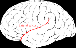
 Lateral sulcus (Middle cerebral veins not visible, but veins run in lateral sulcus.) Lateral sulcus (Middle cerebral veins not visible, but veins run in lateral sulcus.) | |
| Details | |
| Drains to | Cavernous sinus, basal vein |
| Artery | Middle cerebral artery |
| Identifiers | |
| Latin | venae mediae cerebri (superficialis et profunda) |
| Anatomical terminology[edit on Wikidata] | |
The middle cerebral veins - the superficial middle cerebral vein and the deep middle cerebral vein - are two veins running along the lateral sulcus. The superficial middle cerebral vein is also known as the superficial Sylvian vein, and the deep middle cerebral vein is also known as the deep Sylvian vein. The lateral sulcus is also known as the Sylvian fissure.
Superficial middle cerebral vein
The superficial middle cerebral vein (superficial Sylvian vein) begins on the lateral surface of the hemisphere. It runs along the lateral sulcus to empty into either the cavernous sinus, or sphenoparietal sinus. It is adherent to the deep surface of the arachnoid mater bridging the lateral sulcus. It drains the adjacent cortex.
Anastomoses
At its posterior extremity, the superficial middle cerebral vein is connected with the superior sagittal sinus via the superior anastomotic vein, and with the transverse sinus via the inferior anastomotic vein.
Deep middle cerebral vein
The deep middle cerebral vein (deep Sylvian vein) receives tributaries from the insula and neighboring gyri, and runs in the lower part of the lateral sulcus.
Additional images
-
Meninges and superficial cerebral veins. Deep dissection. Superior view.
-
 Base of brain. (Lateral fissure visible at top left.)
-
 Sagittal section of the skull, showing the sinuses of the dura. (Cerebral veins labeled at center left.)
References
- ^
 One or more of the preceding sentences incorporates text in the public domain from page 652 of the 20th edition of Gray's Anatomy (1918)
- ^ Sinnatamby, Chummy (2011). Last's Anatomy (12th ed.). p. 473. ISBN 978-0-7295-3752-0.
-
 One or more of the preceding sentences incorporates text in the public domain from page 653 of the 20th edition of Gray's Anatomy (1918)
| Veins of the head and neck | |||||||||||||||||||||||
|---|---|---|---|---|---|---|---|---|---|---|---|---|---|---|---|---|---|---|---|---|---|---|---|
| External jugular |
| ||||||||||||||||||||||
| Internal jugular |
| ||||||||||||||||||||||
| Brachiocephalic |
| ||||||||||||||||||||||
This cardiovascular system article is a stub. You can help Misplaced Pages by expanding it. |