| Trevor disease | |
|---|---|
| Other names | Dysplasia epiphysealis hemimelica |
 | |
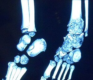
| Trevor disease in a nine-year-old girl: talus | |
| Specialty | Medical genetics |
Trevor disease, also known as dysplasia epiphysealis hemimelica and Trevor's disease, is a congenital bone developmental disorder. There is 1 case per million population. The condition is three times more common in males than in females.
Presentation

This disorder is rare, and is characterised by an asymmetrical limb deformity due to localized overgrowth of cartilage, histologically resembling osteochondroma. It is believed to affect the limb bud in early fetal life. The condition occurs mostly in the ankle or knee region and it is always confined to a single limb. This usually involves only the lower extremities and on medial side of the epiphysis. It is named after researcher David Trevor.
Diagnosis
Differential diagnosis
Trevor disease can often mimic posttraumatic osseous fragments, synovial chondromatosis, ostechondroma, or anterior spur of ankle. It is not possible to distinguish DEH from osteochondroma on the basis of histopathology alone. Special molecular tests of the genes EXT1, EXT2 are used for the analysis of genetic expressions. These are within normal ranges in DEH, while they are lower in ostechondroma (owing to a mutation). These tests are expensive and the diagnosis is often made on clinical and radiological findings. Synovial chondromatosis occurs in a much older age group and can be ruled out on this basis.
Treatment
Most reported cases of DEH in the literature have been treated surgically, usually with excision of the mass, as well as by the correction of any deformity, while preserving the integrity of the affected joint as much as possible.
History
Trevor disease was first described by the French surgeon Albert Mouchet and J. Belot in 1926. In 1956, the name "dysplasia epiphysealis hemimelica" was proposed by Fairbank. The usual symptoms are the appearance of an osseous protuberance, on one side of the knee, ankle or foot joint which gradually increases Radiologically, the condition shows a nonuniformity of growth and multiple unconnected ossification centers around the epiphyses.
See also
References
- ^ www.whonamedit.com
- ^ Gökkus, K; Aydin, AT (2014). "Dysplasia epiphysealis hemimelica: a diagnostic dilemma for orthopedic surgeons and a nightmare for parents". Journal of Postgraduate Medicine. 60 (1): 1–2. doi:10.4103/0022-3859.128794. PMID 24625930.
- Yih-Huie, Lin; Yi-Jiun, Chou; Lee-Ren, Yeh; Clement, K. H.; Chen, Clement K. H.; Chen, Huay-Ban Pan; Chien-Fang, Yang (2001). "Dysplasia Epiphysealis Hemimelica or Trevor's Disease: A Case Report" (PDF). Journal of Radiological Science. 26 (5): 215–20.
- Glick, Rachel; Khaldi, Lubna; Ptaszynski, Konrad; Steiner, German C. (2007). "Dysplasia epiphysealis hemimelica (Trevor disease): a rare developmental disorder of bone mimicking osteochondroma of long bones". Human Pathology. 38 (8): 1265–72. doi:10.1016/j.humpath.2007.01.017. PMID 17490719.
- Murphey, Mark D.; Choi, James J.; Kransdorf, Mark J.; Flemming, Donald J.; Gannon, Frances H. (2000). "Imaging of osteochondroma: variants and complications with radiologic-pathologic correlation". Radiographics. 20 (5): 1407–34. doi:10.1148/radiographics.20.5.g00se171407. PMID 10992031.
- ^ Gokkus, Kemal; Aydin, Ahmet Turan; Uyan, Ayca; Cengiz, Menekse (2011). "Dysplasia epiphysealis hemimelica of the ankle joint: a case report" (PDF). Journal of Orthopaedic Surgery. 19 (2): 254–6. doi:10.1177/230949901101900227. PMID 21857058. S2CID 45805741.
- Gökkuş, Kemal; Aydın, Ahmet Turan; Sagtas, Ergin (2012). "Trevor's disease: mimicking anterior ankle impingement syndrome: case report". Knee Surgery, Sports Traumatology, Arthroscopy. 20 (9): 1875–8. doi:10.1007/s00167-011-1836-y. PMID 22198357. S2CID 23699201.
- ^ Fairbank, TJ (1956). "Dysplasia epiphysialis hemimelica (tarso-ephiphysial aclasis)". The Journal of Bone and Joint Surgery. British Volume. 38-B (1): 237–57. doi:10.1302/0301-620X.38B1.237. PMID 13295331.
- Dorfman, H. D.; Vanel, D.; Czerniak, B.; Park, Y. K.; Kotz, R.; Unni, K. K. (2002). "WHO classification of tumours of bone: Introduction". In Fletcher, Christopher D. M.; Unni, K. Krishnan; Mertens, Fredrik (eds.). Pathology and Genetics of Tumours of Soft Tissue and Bone. IARC. pp. 227–30. ISBN 978-92-832-2413-6.
- Gökkuş, Kemal; Aydın, Ahmet Turan (2010). "Talus ostekondromu ve talus tutulumlu displazi epifiziyalis hemimelika ayırımı, zor bir ikilem" [Differential diagnosis of osteochondroma of talus and talus located dysplasia epiphysealis hemimelica, a diagnostic dilemma] (PDF). Eklem Hastalıkları ve Cerrahisi (in Turkish). 21 (3): 182. PMID 21067502.
- Arkader, Alexandre; Friedman, Jared E; Moroz, Leslie; Dormans, John P (2007). "Acetabular dysplasia with hip subluxation in Trevor's disease of the hip". Clinical Orthopaedics and Related Research. 457: 247–52. doi:10.1097/BLO.0b013e31802ea479. PMID 17146363.
- Maylack, Fallon H.; Manske, Paul R.; Strecker, William B. (1988). "Dysplasia epiphysealis hemimelica at the metacarpophalangeal joint". The Journal of Hand Surgery. 13 (6): 916–20. doi:10.1016/0363-5023(88)90270-5. PMID 3225418.
- Dysplasia Epiphysealis Hemimelica at eMedicine
- Campanacci, Mario (1999). "Hemimelic Epiphyseal Dysplasia". Bone and Soft Tissue Tumors: Clinical Features, Imaging, Pathology and Treatment. pp. 207–211. doi:10.1007/978-3-7091-3846-5_11. ISBN 978-3-7091-3846-5.
External links
| Classification | D |
|---|---|
| External resources |