| Revision as of 15:32, 9 April 2014 editAnthonyhcole (talk | contribs)Extended confirmed users, New page reviewers, Pending changes reviewers39,868 edits →Development of the operculum: Section headings should not refer redundantly to the subject of the article; per Manual of style.← Previous edit |
Revision as of 16:53, 14 April 2014 edit undoIztwoz (talk | contribs)Extended confirmed users, New page reviewers74,456 editsm →Development: typo grammNext edit → |
| (11 intermediate revisions by the same user not shown) |
| Line 1: |
Line 1: |
|
|
|
|
{{Merge to|Parietal_operculum|discuss=Talk:Parietal_operculum#Merger proposal|date=January 2014}} |
|
|
{{Infobox Brain| |
|
{{Infobox Brain| |
|
Name = Operculum (brain) | |
|
Name = Operculum (brain) | |
| Line 20: |
Line 20: |
|
DorlandsSuf = 12592993 | |
|
DorlandsSuf = 12592993 | |
|
}} |
|
}} |
|
In human brain anatomy, '''operculum''' (Latin, meaning "little lid") may refer to the parts of the ], ] and ] lobes that cover the ] (the '''insular operculum'''), or the part of the ] surrounded by the ] (the '''occipital operculum'''). The insular operculum on the ] and ] ] (on either side of the ]) is known as the Rolandic operculum.<ref name="TonkonogyPuente2009">{{cite book|author1=Joseph M. Tonkonogy|author2=Antonio E. Puente|title=Localization of Clinical Syndromes in Neuropsychology and Neuroscience|url=http://books.google.com/books?id=t2Y4gv9d3l0C&pg=PA392|accessdate=12 October 2012|date=23 January 2009|publisher=Springer Publishing Company|isbn=978-0-8261-1967-4|page=392}}</ref> |
|
In ] anatomy, an '''operculum''' (Latin, meaning "little lid") (pl. '''opercula'''), may refer to a part of the ], ] or ] that together cover the ] as the opercula of insula.<ref>Dorlands Illustrated Medical Dictionary 32nd edition 2012 Page 1328 ISBN 978-1-4160-6257-8</ref>It can also refer to the part of the ] surrounded by the '''occipital operculum'''. The insular opercula lie on the ] and ] ] (on either side of the ]).<ref name="TonkonogyPuente2009">{{cite book|author1=Joseph M. Tonkonogy|author2=Antonio E. Puente|title=Localization of Clinical Syndromes in Neuropsychology and Neuroscience|url=http://books.google.com/books?id=t2Y4gv9d3l0C&pg=PA392|accessdate=12 October 2012|date=23 January 2009|publisher=Springer Publishing Company|isbn=978-0-8261-1967-4|page=392}}</ref> |
|
|
|
|
|
The part of the parietal operculum that forms the ceiling of the lateral sulcus functions as the secondary somatosensory cortex. |
|
|
|
|
|
==Albert Einstein's brain== |
|
|
Contrary to the literature, Einstein’s brain is not spherical and has non-confluent Sylvian and inferior postcentral sulci.<ref>"The cerebral cortex of Albert Einstein: a description and preliminary analysis of unpublished photographs". Brain (Oxford University Press). Retrieved 1 December 2012</ref> |
|
|
|
|
|
Opinions differ on whether Einstein’s brain possessed parietal opercula. Falk, et al. claim the brain does have parietal opercula while Witelson et al. claim it does not.<ref>https://www.ncbi.nlm.nih.gov/pubmed/10382713?dopt=Abstract</ref> |
|
|
|
|
|
Einstein's lower parietal lobe (which is responsible for mathematical thought, visuospatial cognition, and imagery of movement) was 15% larger than average. |
|
|
|
|
|
== Development == |
|
== Development == |
|
Normally, the insular operculum begins to develop by 20 to 22 weeks of pregnancy. At 14 to 16 weeks of pregnancy, the insula begins to ] from the surface of the immature cerebrum of the brain, until by the full term of the mother's pregnancy, the operculum completely covers the insula.<ref>Larroche JC. (1977) "Development of the central nervous system". ''Developmental Pathology of the Neonate''. Amsterdam, the Netherlands: ''Excerpta Medica''; 1977:319–327, cited as Note 3 of |
|
Normally, the insular opercula begin to develop between the 20th and the 22nd weeks of pregnancy. At weeks 14 to 16 of ], the insula begins to ] from the surface of the immature cerebrum of the brain, until at ], the opercula completely cover the insula.<ref>Larroche JC. (1977) "Development of the central nervous system". ''Developmental Pathology of the Neonate''. Amsterdam, the Netherlands: ''Excerpta Medica''; 1977:319–327, cited as Note 3 of |
|
*Cheng-Yu Chen, Robert A. Zimmerman, Scott Faro, Beth Parrish, Zhiyue Wang, Larissa T. Bilaniuk, and Ting-Ywan Chou (1996), "MR of the Cerebral Operculum: Abnormal Opercular Formation in Infants and Children" , ''AJNR Am J Neuroradiol'' '''17''':1303–1311, August 1996 |
|
*Cheng-Yu Chen, Robert A. Zimmerman, Scott Faro, Beth Parrish, Zhiyue Wang, Larissa T. Bilaniuk, and Ting-Ywan Chou (1996), "MR of the Cerebral Operculum: Abnormal Opercular Formation in Infants and Children" , ''AJNR Am J Neuroradiol'' '''17''':1303–1311, August 1996 |
|
</ref> |
|
</ref> |
| Line 41: |
Line 50: |
|
] |
|
] |
|
] |
|
] |
|
|
|
|
|
|
|
|
{{neuroscience-stub}} |
|
{{neuroscience-stub}} |
The part of the parietal operculum that forms the ceiling of the lateral sulcus functions as the secondary somatosensory cortex.
Contrary to the literature, Einstein’s brain is not spherical and has non-confluent Sylvian and inferior postcentral sulci.
Opinions differ on whether Einstein’s brain possessed parietal opercula. Falk, et al. claim the brain does have parietal opercula while Witelson et al. claim it does not.
Einstein's lower parietal lobe (which is responsible for mathematical thought, visuospatial cognition, and imagery of movement) was 15% larger than average.
Normally, the insular opercula begin to develop between the 20th and the 22nd weeks of pregnancy. At weeks 14 to 16 of fetal development, the insula begins to invaginate from the surface of the immature cerebrum of the brain, until at full term, the opercula completely cover the insula.
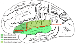
 Operculum
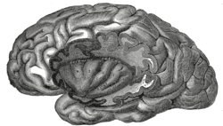
 The insula of the left side, exposed by removing the operculum