This is an old revision of this page, as edited by Opabinia regalis (talk | contribs) at 02:02, 6 January 2007 (formatting). The present address (URL) is a permanent link to this revision, which may differ significantly from the current revision.
Revision as of 02:02, 6 January 2007 by Opabinia regalis (talk | contribs) (formatting)(diff) ← Previous revision | Latest revision (diff) | Newer revision → (diff)

Proteasomes are large protein complexes inside all eukaryotes and archaea, as well as some bacteria. In eukaryotes, they are located in the nucleus and the cytoplasm. The main function of the proteasome is to degrade unneeded or damaged proteins by proteolysis, a chemical reaction that breaks peptide bonds. Enzymes that carry out such reactions are called proteases. Proteasomes are a major mechanism by which cells regulate the concentration of particular proteins and degrade misfolded proteins. The degradation process yields peptides of about 7–8 amino acids long, which can then be further degraded into amino acids and used in synthesizing new proteins. Proteins are tagged for degradation by a small protein called ubiquitin. The tagging reaction is catalyzed by enzymes called ubiquitin ligases. Once a protein is tagged with a single ubiquitin molecule, this is a signal to other ligases to attach additional ubiquitin molecules. The result is a polyubiquitin chain, which is recognized by the proteasome and allows it to bind and degrade the tagged protein.
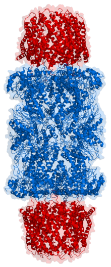
Structurally, the proteasome is a large barrel-like complex containing a "core" of four stacked rings around a central pore. Each ring is in turn composed of seven individual proteins. The inner two rings are made from seven β subunits that contain the protease active sites. These sites are located on the interior surface of the rings, so that the target protein must enter the central pore before it is degraded. The outer two rings each contain seven α subunits whose function is to maintain a "gate" through which proteins enter the barrel. These α subunits are controlled by binding to "cap" structures or regulatory particles that recognize polyubiquitin tags attached to protein substrates and initiate the degradation process. The overall system of ubiquitination and proteasomal degradation is known as the ubiquitin-proteasome system.
The proteasomal degradation pathway is essential for many cellular processes, including the cell cycle, the regulation of gene expression, and responses to oxidative stress. The importance of proteolytic degradation inside cells and the role of ubiquitin in proteolytic pathways was acknowledged in the awarding of the 2004 Nobel Prize in Chemistry to Aaron Ciechanover, Avram Hershko and Irwin Rose.
Discovery
Before the discovery of the ubiquitin proteasome system, protein degradation in cells was thought to rely mainly on lysosomes, membrane-bound organelles with acidic and protease-filled interiors that can degrade and then recycle exogenous proteins and aged or damaged organelles. However, work on ATP-dependent protein degradation in reticulocytes, which lack lysosomes, suggested the presence of a second intracellular degradation mechanism. This was shown in 1978 to be composed of several distinct protein chains, a novelty among proteases at the time. Later work on modification of histones led to the identification of an unexpected covalent modification of the histone protein by a branched bond between a histone lysine residue and the N-terminal glycine residue of the protein ubiquitin, which had no known function. It was then discovered that a previously-identified protein associated with proteolytic degradation, known as ATP-dependent proteolysis factor 1 (APF-1), was the same protein as ubiquitin.
Much of the early work leading up to the discovery of the ubiquitin proteasome system occurred in the late 1970s and early 1980s at the Technion in the laboratory of Avram Hershko, where Aaron Ciechanover worked as a graduate student. Hershko's year-long sabbatical in the laboratory of Irwin Rose at the Fox Chase Cancer Center provided key conceptual insights, though Rose later downplayed his role in the discovery. The three shared the 2004 Nobel Prize in Chemistry for their work in discovering this system.
Although electron microscopy data revealing the stacked-ring structure of the proteasome became available in the mid-1980's, the first structure of the proteasome core particle was not solved by X-ray crystallography until 1994. As of 2006, no structure has been solved of the core particle in complex with the most common form of regulatory cap.
Structure and organization
Template:ImageStackRight The proteasome subcomponents are often referred to by their Svedberg sedimentation coefficient (denoted S). The most common form of the proteasome is known as the 26S proteasome, which is about 2000 kilodaltons (kDa) in molecular mass and contains one 20S core particle structure and two 19S regulatory caps. The core is hollow and provides an enclosed cavity in which proteins are degraded; openings at the two ends of the core allow the target protein to enter. Each end of the core particle associates with a 19S regulatory subunit that contains multiple ATPase active sites and ubiquitin binding sites; it is this structure that recognizes polyubiquitinated proteins and transfers them to the catalytic core. An alternative form of regulatory subunit called the 11S particle can associate with the core in essentially the same manner as the 19S particle; the 11S may play a role in degradation of foreign peptides such as those produced after infection by a virus.
20S core particle
The number and diversity of subunits represented in the 20S core particle depends on the organism; the number of distinct and specialized subunits is larger in multicellular than unicellular organisms and larger in eukaryotes than in prokaryotes. All 20S particles consist of four layers of a heptameric ring structures that are themselves composed of two different types of subunits; α subunits are structural in nature, while β subunits are predominantly catalytic. The outer two rings in the stack consist of seven α subunits each, which serve as docking domains for the regulatory particles and form an exterior gate blocking unregulated access to the interior. The inner two rings each consist of seven β subunits and contain the protease active sites that perform the proteolysis reactions. The size of the proteasome is relatively conserved and is about 150 angstroms (Å) by 115 Å. The interior chamber is at most 53 Å wide, though the entrance can be as narrow as 13 Å, suggesting that substrate proteins must be at least partially unfolded to enter.
In archaea such as Thermoplasma acidophilum, all the α and all the β subunits are identical, while eukaryotic proteasomes such as those in yeast contain seven distinct types of each subunit. In mammals, the β1, β2, and β5 subunits are catalytic; although they share a common mechanism, they have three distinct substrate specificities considered chymotrypsin-like, trypsin-like, and peptidyl-glutamyl peptide-hydrolyzing (PHGH). Alternative β forms denoted β1i, β2i, and β5i can be expressed in hematopoietic cells in response to exposure to pro-inflammatory signals such as cytokines, in particular, interferon gamma. The proteasome assembled with these alternative subunits is known as the immunoproteasome, whose substrate specificity is altered relative to the normal proteasome.
19S regulatory particle
The 19S particle in eukaryotes consists of 19 individual proteins and is divisible into two subunits, a 10-protein base that binds directly to the α ring of the 20S core particle, and a 9-protein lid where polyubiquitin is bound. Six of the ten base proteins have ATPase activity. The association of the 19S and 20S particles requires the binding of ATP to the 19S ATP-binding sites. ATP hydrolysis is required for the assembled complex to degrade a folded and ubiquitinated protein, although it is not yet clear whether that energy is used mainly for substrate unfolding, opening of the core channel, or some combination of processes. As of 2006, the structure of the 26S proteasome has not been solved, although the 19S and 11S particles are believed to bind in a similar way to the α rings of the 20S core particle.
Individual components of the 19S particle have their own regulatory roles. Gankyrin, a recently identified oncoprotein, is one of the 19S subcomponents that also tightly binds the cyclin-dependent kinase CDK4 and plays a key role in recognizing ubiquitinated p53, via its affinity for the ubiquitin ligase MDM2. Gankyrin is anti-apoptotic and has been shown to be overexpressed in some tumor cell types such as hepatocellular carcinoma.
11S regulatory particle
20S proteasomes can also associate with a second type of regulatory particle, a heptameric structure that does not contain any ATPases and can promote the degradation of short peptides, but not of complete proteins. Presumably this is because the complex cannot unfold larger substrates. This structure is also known as PA28 or REG. The mechanisms by which it binds to the core particle through the C-terminal tails of its subunits and induces α-ring conformational changes to open the 20S gate suggest a similar mechanism for the 19S particle. The expression of the 11S particle is induced by interferon gamma and is responsible, in conjunction with the immunoproteasome β subunits, for the generation of peptides that bind to the major histocompatibility complex.
Assembly
The assembly of the proteasome is a complex process due to the number of subunits that must associate to form an active complex. The β subunits are synthesized with N-terminal "propeptides" that are post-translationally modified during the assembly of the 20S particle to expose the proteolytic active site. The 20S particle is assembled from two half-proteasomes, each of which consists of a seven-membered pro-β ring attached to a seven-membered α ring. The association of the β rings of the two half-proteasomes triggers threonine-dependent autolysis of the propeptides to expose the active site. These β interactions are mediated mainly by salt bridges and hydrophobic interactions between conserved alpha helices whose disruption by mutation damages the proteasome's ability to assemble. The assembly of the half-proteasomes, in turn, is initiated by the assembly of the α subunits into their heptameric ring, forming a template for the association of the corresponding pro-β ring. The assembly of α subunits has not been characterized.
In general, less is known about the assembly and maturation of the 19S regulatory particles. They are believed to assemble as two distinct subcomponents, the ATPase-containing base and the ubiquitin-recognizing lid. The six ATPases in the base may assemble in a pairwise manner mediated by coiled-coil interactions. The order in which the nineteen subunits of the regulatory particle are bound is a likely regulatory mechanism that prevents exposure of the active site before assembly is complete.
The protein degradation process

Ubiquitination and targeting
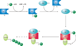
Proteins are targeted for degradation by the proteasome by covalent modification of a lysine residue that requires the coordinated reactions of three enzymes. In the first step, a ubiquitin-activating enzyme (known as E1) hydrolyzes ATP and adenylates a ubiquitin molecule. This is then transferred to E1's active-site cysteine residue in concert with the adenylation of a second ubiquitin. This adenylated ubiquitin is then transferred to a cysteine of a second enzyme, ubiquitin-conjugating enzyme (E2). Lastly, a member of a highly-diverse class of enzymes known as ubiquitin ligases (E3) recognize the specific protein to be ubiquitinated and catalyze the transfer of ubiquitin from E2 to this target protein. A target protein must be labelled with at least four ubiquitin monomers (in the form of a polyubiquitin chain) before it is recognized by the proteasome lid. It is therefore the E3 that confers substrate specificity to this system. The number of E1, E2, and E3 proteins expressed depends on the organism and cell type, but there are many different E3 enzymes present in humans, indicating that there are a huge number of targets for the ubiquitin proteasome system.
The mechanism by which a polyubiquitinated protein is targeted to the proteasome is not fully understood. Ubiquitin-receptor proteins have an N-terminal ubiquitin-like (UBL) domain and one or more ubiquitin-associated (UBA) domains. The UBL domains are recognized by the 19S proteasome caps and the UBA domains bind ubiquitin via three-helix bundles. These receptor proteins may escort polyubiquitinated proteins to the proteasome, though the specifics of this interaction and its regulation are unclear.
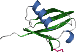
The ubiquitin protein itself is 76 amino acids long and was named due to its ubiquitous nature, as it has a highly conserved sequence and is found in all known organisms. The genes encoding ubiquitin in eukaryotes are arranged in tandem repeats, possibly due to the heavy transcription demands on these genes to produce enough ubiquitin for the cell. It has been proposed that ubiquitin is the slowest-evolving protein identified to date.

Unfolding and translocation
After a protein has been ubiquitinated, it is recognized by the 19S regulatory particle in an ATP-dependent binding step. The substrate protein must then enter the interior of the 20S particle to come in contact with the proteolytic active sites. Because the 20S particle's central channel is narrow and gated by the N-terminal tails of the α ring subunits, the substrates must be at least partially unfolded before they enter the core. The passage of the unfolded substrate into the core is called translocation and necessarily occurs after deubiquitination. However, the order in which substrates are deubiquitinated and unfolded is not yet clear. Which of these processes is the rate-limiting step in the overall proteolysis reaction depends on the specific substrate; for some proteins, the unfolding process is rate-limiting, while deubiquitination is the slowest step for other proteins. The extent to which substrates must be unfolded before translocation is not known, but substantial tertiary structure, and in particular nonlocal interactions such as disulfide bonds, are sufficient to inhibit degradation.
The gate formed by the α subunits prevents peptides longer than about four residues from entering the interior of the 20S particle. The ATP molecules bound before the initial recognition step are hydrolyzed before translocation, although there is disagreement whether the energy is needed for substrate unfolding or for gate opening. The assembled 26S proteasome can degrade unfolded proteins in the presence of a non-hydrolyzable ATP analog, but cannot degrade folded proteins, indicating that energy from ATP hydrolysis is used for substrate unfolding. Passage of the unfolded substrate through the opened gate occurs via facilitated diffusion if the 19S cap is in the ATP-bound state.
The mechanism for unfolding of globular proteins is necessarily general, but somewhat dependent on the amino acid sequence. Long sequences of alternating glycine and alanine have been shown to inhibit substrate unfolding decreasing the efficiency of proteasomal degradation; this results in the release of partially degraded byproducts, possibly due to the decoupling of the ATP hydrolysis and unfolding steps. Such glycine-alanine repeats are also found in nature, for example in silk fibroin; in particular, certain Epstein-Barr virus gene products bearing this sequence can stall the proteasome, helping the virus propagate by preventing antigen presentation on the major histocompatibility complex.
Proteolysis
The mechanism of proteolyisi by the β subunits of the 20S core particle is through a threonine-dependent nucleophilic attack. This mechanism may depend on an associated water molecule for deprotonation of the reactive threonine hydroxyl. Degradation occurs within the central chamber formed by the association of the two β rings and normally does not release partially degraded products, instead reducing the substrate to short polypeptides typically 7–9 residues long, though they can range from 4 to 25 residues depending on the organism and substrate. The biochemical mechanism that determines product length is not fully characterized. Although the three catalytic β subunits share a common mechanism, they have slightly different substrate specificities, which are considered chymotrypsin-like, trypsin-like, and peptidyl-glutamyl peptide-hydrolyzing (PHGH)-like. These variations in specificity are the result of interatomic contacts with local residues near the active sites of each subunit. Each catalytic β subunit also possesses a conserved lysine residue required for proteolysis.
Although the proteasome normally produces very short peptide fragments, in some cases these products are themselves biologically active and functional molecules. Certain transcription factors, including one component of the mammalian complex NF-κB, are synthesized as inactive precursors whose ubiquitination and subsequent proteasomal degradation converts them to an active form. Such activity requires the proteasome to cleave the substrate protein internally: rather than processively degrading it from one terminus. It has been suggested that long loops on these proteins' surfaces serve as the proteasomal substrates and enter the central cavity, while the majority of the protein remains outside. Similar effects have been observed in yeast proteins; this mechanism of selective degradation is known as regulated ubiquitin/proteasome dependent processing (RUP).
Ubiquitin-independent degradation
Although most proteasomal substrates must be ubiquitinated before being degraded, there are some exceptions to this general rule, especially when the proteasome plays a normal role in the post-translational processing of the protein. The proteasomal activation of NF-κB by processing p105 into p50 via internal proteolysis is one major example. Some proteins that are hypothesized to be unstable due to intrinsically unstructured regions, are degraded in a ubiquitin-independent manner. The most well-known example of a ubiquitin-independent proteasome substrate is the enzyme ornithine decarboxylase. Ubiquitin-independent mechanisms targeting key cell cycle regulators such as p53 have also been reported, although p53 is also subject to ubiquitin-dependent degradation. Finally, structurally-abnormal, misfolded, or highly oxidized proteins are also subject to ubiquitin-independent and 19S-independent degradation under conditions of cellular stress.
Evolution

The 20S proteasome is both ubiquitous and essential in eukaryotes. Some prokaryotes, including many archaea and the bacterial order Actinomycetales also share homologs of the 20S proteasome, while most bacteria possess heat shock genes hslV and hslU, whose gene products are a multimeric protease arranged in a two-layered ring and an ATPase. The hslV protein has been hypothesized to resemble the likely ancestor of the 20S proteasome. HslV is generally not essential in bacteria, and not all bacteria possess it, while some protists possess both the 20S and the hslV systems.
Sequence analysis suggests that the catalytic β subunits diverged earlier in evolution than the predominantly-structural α subunits. In bacteria that express a 20S proteasome, the β subunits have high sequence identity to archaeal and eukaryotic β subunits, while the α sequence identity is much lower. The presence of 20S proteasomes in bacteria may result from lateral gene transfer, while the diversification of subunits among eukaryotes is ascribed to multiple gene duplication events.
Cell cycle control
Cell cycle progression is controlled by ordered action of cyclin-dependent kinases (CDKs), activated by specific cyclins that demarcate phases of the cell cycle. Mitotic cyclins, which persist in the cell for only a few minutes, have one of the shortest life spans of all intracellular proteins. After a CDK-cyclin complex has performed its function, the associated cyclin is polyubiquitinated and destroyed by the proteasome, which provides directionality for the cell cycle. In particular, exit from mitosis requires the proteasome-dependent dissociation of the regulatory component cyclin B from the mitosis promoting factor complex. In vertebrate cells, "slippage" through the mitotic checkpoint leading to premature M phase exit can occur despite the delay of this exit by the spindle checkpoint.
Earlier cell cycle checkpoints such a post-restriction point check between G1 phase and S phase similarly involve proteasomal degradation of cyclin A, whose ubiquitination is promoted by the anaphase promoting complex (APC), an E3 ubiquitin ligase. The APC and the Skp1/Cul1/F-box protein complex (SCF complex) are the two key regulators of cyclin degradation and checkpoint control; the SCF itself is regulated by the APC via ubiquitination of the Skp1 component, which prevents SCF activity before the G1-S transition.
Regulation of plant growth
In plants, signaling by auxins, or phytohormones that order the direction and tropism of plant growth, induces the targeting of a class of transcription factor repressors known as Aux/IAA proteins for proteasomal degradation. These proteins are ubiquitinated by SCFTIR1, or SCF in complex with the auxin receptor TIR1. Degradation of Aux/IAA proteins derepresses transcription factors in the auxin-response factor (ARF) family and induces ARF-directed gene expression. The cellular consequences of ARF activation depend on the plant type and developmental stage, but are involved in directing growth in roots and leaf veins. The specific response to ARF derepression is thought to be mediated by specificity in the pairing of individual ARF and Aux/IAA proteins.
Apoptosis
Both internal and external signals can lead to the induction of apoptosis, or programmed cell death. The resulting deconstruction of cellular components is primarily carried out by specialized proteases known as caspases, but the proteasome also plays important and diverse roles in the apoptotic process. The involvement of the proteasome in this process is indicated by both the increase in protein ubiquitination, and of E1, E2, and E3 enzymes that is observed well in advance of apoptosis, During apoptosis, proteasomes localized to the nucleus have also been observed to translocate to outer membrane blebs characteristic of apoptosis.
Proteasome inhibition has different effects on apoptosis induction in different cell types. The proteasome is not generally required for apoptosis, although inhibiting it is pro-apoptotic in most cell types that have been studied. However, some cell lines—in particular, primary cultures of quiescent and differentiated cells such as thymocytes and neurons—are prevented from undergoing apoptosis on exposure to proteasome inhibitors. The mechanism for this effect is not clear, but is hypothesized to be specific to cells in quiescent states, or to result from the differential activity of the pro-apoptotic kinase JNK. The ability of proteasome inhibitors to induce apoptosis in rapidly dividing cells has been exploited in several recently developed chemotherapy agents.
Response to cellular stress
In response to cellular stresses - such as infection, heat shock, or oxidative damage - heat shock proteins are expressed that identify misfolded or unfolded proteins and target them for proteasomal degradation. Both Hsp27 and Hsp90—chaperone proteins have been implicated in increasing the activity of the ubiquitin-proteasome system, though they are not direct participants in the process. Hsp70, on the other hand, binds exposed hydrophobic patches on the surface of misfolded proteins and recruits E3 ubiquitin ligases such as CHIP to tag the proteins for proteasomal degradation. The CHIP protein (carboxyl terminus of Hsp70-interacting protein) is itself regulated via inhibition of interactions between the E3 enzyme CHIP and its E2 binding partner.
Similar mechanisms exist to promote the degradation of oxidatively-damaged proteins via the proteasome system. In particular, proteasomes localized to the nucleus are regulated by PARP and actively degrade inappropriately oxidized histones. Oxidized proteins, which often form large amorphous aggregates in the cell, can be degraded directly by the 20S core particle without the 19S regulatory cap and do not require ATP hydrolysis or tagging with ubiquitin. However, high levels of oxidative damage increases the degree of cross-linking between protein fragments, rendering the aggregates resistant to proteolysis. Larger numbers and sizes of such highly oxidized aggregates are associated with aging.
Impaired proteasomal activity has been suggested as an explanation for some of the late-onset neurodegenerative diseases that share aggregation of misfolded proteins as a common feature, such as Parkinson's disease and Alzheimer's disease. In these diseases large insoluble aggregates of misfolded proteins can form and then result in neurotoxicity, through mechanisms that are not yet well understood. Decreased proteasome activity has been suggested as a cause of aggregation and Lewy body formation in Parkinson's. This hypothesis is supported by the observation that yeast models of Parkinson's are more susceptible to toxicity from alpha-synuclein, the major protein component of Lewy bodies, under conditions of low proteasome activity.
Role in the immune system
The proteasome plays a straightforward but critical role in the function of the adaptive immune system. Peptide antigens are displayed by the major histocompatibility complex class I (MHC) proteins on the surface of antigen-presenting cells. These peptides are products of proteasomal degradation of proteins originated by the invading pathogen. Although constitutively-expressed proteasomes can participate in this process, a specialized complex composed of proteins whose expression is induced by interferon gamma produces peptides of the optimal size and composition for MHC binding. These proteins whose expression increases during the immune response include the 11S regulatory particle, whose main known biological role is regulating the production of MHC ligands, and specialized β subunits called β1i, β2i, and β5i with altered substrate specificity. The complex formed with the specialized β subunits is known as the immunoproteasome.
The strength of MHC class I ligand binding is dependent on the composition of the ligand C-terminus, as peptides bind by hydrogen bonding and by close contacts with a region called the "B pocket" on the MHC surface. Optimal residues for the C-terminal end of these peptides are leucine and valine. The N-terminal residues, particularly the second residue in the peptide, also play a key role in determining binding affinity. The immunoproteasome complex generates the correct C-terminal tails; later processing of the products by interferon gamma-induced aminopeptidases trims the N-termini for optimal MHC ligand production.
Due to its role in generating the activated form of NF-κB, an anti-apoptotic and pro-inflammatory regulator of cytokine expression, proteasomal activity has been linked to inflammatory and autoimmune diseases. Increased levels of proteasome activity correlate with disease activity and have been implicated in autoimmune diseases including systemic lupus erythematosus and rheumatoid arthritis.
Proteasome inhibitors


Proteasome inhibitors have effective anti-tumor activity in cell culture, inducing apoptosis by disrupting the regulated degradation of pro-growth cell cycle proteins. This approach of selectively-inducing apoptosis in tumor cells has proven effective in animal models and human trials. Bortezomib, a molecule developed by Millennium Pharmaceuticals and marketed as Velcade, is the first proteasome inhibitor to reach clinical use as a chemotherapy agent. Bortezomib is used in the treatment of multiple myeloma. Notably, multiple myeloma has been observed to result in increased proteasome levels in blood serum that decrease to normal levels in response to successful chemotherapy. Studies in animals have indicated that bortezomib may also have clinically-significant effects in pancreatic cancer. Preclinical and early clinical studies have been started to examine bortezomib's effectiveness in treating other B-cell-related cancers, particularly some types of non-Hodgkin's lymphoma.
The molecule ritonavir, marketed as Norvir, was developed as a protease inhibitor and used to target HIV infection. However, it has been shown to inhibit proteasomes as well as free proteases; specifically, the chymotrypsin-like activity of the proteasome is inhibited by ritonavir, while the trypsin-like activity is somewhat enhanced. Studies in animal models suggest that ritonavir may have inhibitory effects on the growth of glioma cells.
Proteasome inhibitors have also shown promise in treating autoimmune diseases in animal models. For example, studies in mice bearing human skin grafts found a reduction in the size of lesions from psoriasis after treatment with a proteasome inhibitor. Inhibitors also show positive effects in rodent models of asthma.
Labeling and inhibition of the proteasome is also of interest in laboratory settings for both in vitro and in vivo study of proteasomal activity in cells. The most commonly used laboratory inhibitor is lactacystin, a natural product synthesized by Streptomyces bacteria. Fluorescent inhibitors have also been developed to specifically label the active sites of the assembled proteasome.
References
- Peters JM, Franke WW, Kleinschmidt JA. (1994) Distinct 19S and 20S subcomplexes of the 26S proteasome and their distribution in the nucleus and the cytoplasm. J Biol Chem, Mar 11;269(10):7709-18. PMID 8125997
- ^ Lodish, H, Berk A, Matsudaira P, Kaiser CA, Krieger M, Scott MP, Zipursky SL, Darnell J. (2004). Molecular Cell Biology, 5th ed., ch.3, pp 66-72. New York: WH Freeman. ISBN 0716743663
- ^ Nobel Prize Committee (2004). "Nobel Prize Awardees in Chemistry, 2004". Retrieved 2006-12-11.
- Ciehanover A, Hod Y, Hershko A. (1978). A heat-stable polypeptide component of an ATP-dependent proteolytic system from reticulocytes. Biochem Biophys Res Commun. 81(4):1100-1105. PMID 666810
- Ciechanover A. (2000). Early work on the ubiquitin proteasome system, an interview with Aaron Ciechanover. Cell Death Differ Sep;12(9):1167-77. PMID 16094393
- Hershko A. (2005). Early work on the ubiquitin proteasome system, an interview with Avram Hershko. Cell Death Differ 12, 1158–1161. PMID 16094391
- Kopp F, Steiner R, Dahlmann B, Kuehn L, Reinauer H. (1986). Size and shape of the multicatalytic proteinase from rat skeletal muscle. Biochim Biophys Acta 872(3):253-60. PMID 3524688
- Löwe J, Stock D, Jap B, Zwickl P, Baumeister W, Huber R. (1995). Crystal structure of the 20S proteasome from the archaeon T. acidophilum at 3.4 Å resolution. Science 268, 533-539. PMID 7725097
- ^ Wang J, Maldonado MA. (2006). The Ubiquitin-Proteasome System and Its Role in Inflammatory and Autoimmune Diseases. Cell Mol Immunol 3(4): 255. PMID 16978533
- ^ Nandi D, Tahiliani P, Kumar A, Chandu D. (2006). The ubiquitin-proteasome system. J Biosci Mar;31(1):137-55. PMID 16595883
- ^ Heinemeyer W, Fischer M, Krimmer T, Stachon U, Wolf DH. (1997). The Active Sites of the Eukaryotic 20S Proteasome and Their Involvement in Subunit Precursor Processing J Biol Chem 272(40):25200-25209. PMID 9312134
- ^ Liu CW, Li X, Thompson D, Wooding K, Chang TL, Tang Z, Yu H, Thomas PJ, DeMartino GN. (2006). ATP binding and ATP hydrolysis play distinct roles in the function of 26S proteasome. Mol Cell Oct 6;24(1):39-50. PMID 17018291
- ^ Lam YA, Lawson TG, Velayutham M, Zweier JL, Pickart CM. (2002). A proteasomal ATPase subunit recognizes the polyubiquitin degradation signal. Nature 416(6882):763-7. PMID 11961560
- ^ Sharon M, Taverner T, Ambroggio XI, Deshaies RJ, Robinson CV. (2006). Structural Organization of the 19S Proteasome Lid: Insights from MS of Intact Complexes. PLoS Biol Aug;4(8):e267. PMID 16869714
- Higashitsuji H, Liu Y, Mayer RJ, Fujita J. (2005). The oncoprotein gankyrin negatively regulates both p53 and RB by enhancing proteasomal degradation. Cell Cycle 4(10):1335-7. PMID 16177571
- Forster A, Masters EI, Whitby FG, Robinson H, Hill CP. (2005). The 1.9 Å Structure of a Proteasome-11S Activator Complex and Implications for Proteasome-PAN/PA700 Interactions. Mol Cell May 27;18(5):589-99. PMID 15916965
- Witt S, Kwon YD, Sharon M, Felderer K, Beuttler M, Robinson CV, Baumeister W, Jap BK. (2006). Proteasome assembly triggers a switch required for active-site maturation. Structure Jul;14(7):1179-88. PMID 16843899
- Kruger E, Kloetzel PM, Enenkel C. (2001). 20S proteasome biogenesis. Biochimie Mar-Apr;83(3-4):289-93. PMID 11295488
- Gorbea C, Taillandier D, Rechsteiner M. (1999). Assembly of the regulatory complex of the 26S proteasome. Mol Biol Rep Apr;26(1-2):15-9. PMID 10363641
- ^ Haas AL, Warms JV, Hershko A, Rose IA. Ubiquitin-activating enzyme: Mechanism and role in protein-ubiquitin conjugation. J Biol Chem. 1982 Mar 10;257(5):2543-8. PMID 6277905 Cite error: The named reference "Haas" was defined multiple times with different content (see the help page).
- Thrower JS, Hoffman L, Rechsteiner M, Pickart CM. (2000). Recognition of the polyubiquitin proteolytic signal. EMBO J 19, 94–102. PMID 10619848
- Risseeuw EP, Daskalchuk TE, Banks TW, Liu E, Cotelesage J, Hellmann H, Estelle M, Somers DE, Crosby WL. Protein interaction analysis of SCF ubiquitin E3 ligase subunits from Arabidopsis. Plant J. 2003 Jun;34(6):753-67. PMID 12795696
- Elsasser S, Finley D. (2005). Delivery of ubiquitinated substrates to protein-unfolding machines. Nat Cell Biol 7(8):742-9. PMID 16056265
- Sharp PM, Li WH. (1987). Ubiquitin genes as a paradigm of concerted evolution of tandem repeats. J Mol Evol 25(1):1432-1432. PMID 3041010
- Zhu Q, Wani G, Wang QE, El-mahdy M, Snapka RM, Wani AA. (2005). Deubiquitination by proteasome is coordinated with substrate translocation for proteolysis in vivo. Exp Cell Res 307(2):436-51. PMID 15950624
- Wenzel T, Baumeister W. (1995). Conformational constraints in protein degradation by the 20S proteasome. Nat Struct Biol 2: 199-204. PMID 7773788
- Smith DM, Kafri G, Cheng Y, Ng D, Walz T, Goldberg AL. (2005). ATP binding to PAN or the 26S ATPases causes association with the 20S proteasome, gate opening, and translocation of unfolded proteins. Mol Cell 20(5):687-98. PMID 16337593
- Smith DM, Benaroudj N, Goldberg A. (2006). Proteasomes and their associated ATPases: a destructive combination. J Struct Biol 156(1):72-83. PMID 16919475
- Hoyt MA, Zich J, Takeuchi J, Zhang M, Govaerts C, Coffino P. (2006). Glycine-alanine repeats impair proper substrate unfolding by the proteasome. EMBO J 25(8):1720-9. PMID 16601692
- ^ Zhang M, Coffino P. (2004). Repeat sequence of Epstein-Barr virus-encoded nuclear antigen 1 protein interrupts proteasome substrate processing. J Biol Chem 279(10):8635-41. PMID 14688254 Cite error: The named reference "Zhang" was defined multiple times with different content (see the help page).
- Voges D, Zwickl P, Baumeister W. (1999). The 26S proteasome: a molecular machine designed for controlled proteolysis. Annu Rev Biochem 68: 1015-1068. PMID 10872471
- ^ Rape M, Jentsch S. (2002). Taking a bite: proteasomal protein processing. Nat Cell Biol 4(5):E113-6. PMID 11988749
- Rape M, Jentsch S. (2004). Productive RUPture: activation of transcription factors by proteasomal processing. Biochim Biophys Acta 1695(1-3):209-13. PMID 15571816
- Asher G, Reuven N, Shaul Y. (2006). 20S proteasomes and protein degradation "by default". Bioessays 28(8):844-9. PMID 16927316
- Asher G, Shaul Y. (2005). p53 proteasomal degradation: poly-ubiquitination is not the whole story. Cell Cycle 4(8):1015-8. PMID 16082197
- ^ Shringarpure R, Grune T, Mehlhase J, Davies KJ. (2003). Ubiquitin conjugation is not required for the degradation of oxidized proteins by proteasome. J Biol Chem 278(1):311-8. PMID 12401807
- ^ Gille C, Goedel A, Schloetelburg C, Preißner R, Kloetzell PM, Gobel UB, Frommell C. (2003). A Comprehensive View on Proteasomal Sequences: Implications for the Evolution of the Proteasome. J Mol Biol 326: 1437–1448. PMID 12595256
- Bochtler M, Ditzel L, Groll M, Hartmann C, Huber R. (1999). The proteasome. Annu. Rev. Biophys. Biomol. Struct. 28: 295–317. PMID 10410804
- Chesnel F, Bazile F, Pascal A, Kubiak JZ. (2006). Cyclin B dissociation from CDK1 precedes its degradation upon MPF inactivation in mitotic extracts of Xenopus laevis embryos. Cell Cycle 5(15):1687-98. PMID 16921258
- Brito DA, Rieder CL. (2006). Mitotic checkpoint slippage in humans occurs via cyclin B destruction in the presence of an active checkpoint. Curr Biol 16(12):1194-200. PMID 16782009
- Havens CG, Ho A, Yoshioka N, Dowdy SF. (2006). Regulation of late G1/S phase transition and APC Cdh1 by reactive oxygen species. Mol Cell Biol 26(12):4701-11. PMID 16738333
- Bashir T, Dorrello NV, Amador V, Guardavaccaro D, Pagano M. (2004). Control of the SCF(Skp2-Cks1) ubiquitin ligase by the APC/C(Cdh1) ubiquitin ligase. Nature 428(6979):190-3. PMID 15014502
- Dharmasiri S, Estelle M. (2002). The role of regulated protein degradation in auxin response. Plant Mol Biol 49(3-4):401-9. PMID 12036263
- Weijers D, Benkova E, Jager KE, Schlereth A, Hamann T, Kientz M, Wilmoth JC, Reed JW, Jurgens G. (2005). Developmental specificity of auxin response by pairs of ARF and Aux/IAA transcriptional regulators. EMBO J 24(10):1874-85. PMID 15889151
- Schwartz LM, Myer A, Kosz L, Engelstein M and Maier C. (1990) Activation of polyubiquitin gene expression during developmentally programmed cell death. Neuron 5: 411-419. PMID 2169771
- Low P, Bussell K, Dawson SP, Billett MA, Mayer RJ and Reynolds SE (1997) Expression of a 26S proteasome ATPase subunit, MS73, in muscles that undergo developmentally programmed cell death, and its control by ecdysteroid hormones in the insect Manduca sexta. FEBS Lett. 400: 345-349. PMID 9009228
- Pitzer F, Dantes A, Fuchs T, Baumeister W, Amsterdam A (1996) Removal of proteasomes from the nucleus and their accumulation in apoptotic blebs during programmed cell death. FEBS Lett. 394: 47-50. PMID 8925925
- ^ Orlowski RZ. (1999). The role of the ubiquitin-proteasome pathway in apoptosis. Cell Death Differ 6: 303-313. PMID 10381632
- Garrido C, Brunet M, Didelot C, Zermati Y, Schmitt E, Kroemer G. (2006). Heat Shock Proteins 27 and 70: Anti-Apoptotic Proteins with Tumorigenic Properties. Cell Cycle 5(22). PMID 17106261
- Park SH, Bolender N, Eisele F, Kostova Z, Takeuchi J, Coffino P, Wolf DH. (2006). The Cytoplasmic Hsp70 Chaperone Machinery Subjects Misfolded and ER Import Incompetent Proteins to Degradation via the Ubiquitin-Proteasome System. Mol Biol Cell Epub. PMID 17065559
- Dai Q, Qian SB, Li HH, McDonough H, Borchers C, Huang D, Takayama S, Younger JM, Ren HY, Cyr DM, Patterson C. (2005). Regulation of the cytoplasmic quality control protein degradation pathway by BAG2. J Biol Chem 280(46):38673-81. PMID 16169850
- Bader N, Grune T. (2006). Protein oxidation and proteolysis. Biol Chem 387(10-11):1351-5. PMID 17081106
- ^ Davies KJ. (2003). Degradation of oxidized proteins by the 20S proteasome. Biochimie 83(3-4):301-10. PMID 11295490 Cite error: The named reference "Davies" was defined multiple times with different content (see the help page).
- McNaught KS, Jackson T, JnoBaptiste R, Kapustin A, Olanow CW. (2006). Proteasomal dysfunction in sporadic Parkinson's disease. Neurology 66(10 Suppl 4):S37-49. PMID 16717251
- Sharma N, Brandis KA, Herrera SK, Johnson BE, Vaidya T, Shrestha R, Debburman SK. (2006). Alpha-Synuclein budding yeast model: toxicity enhanced by impaired proteasome and oxidative stress. J Mol Neurosci 28(2):161-78. PMID 16679556
- Sliz P, Michielin O, Cerottini JC, Luescher I, Romero P, Karplus M, Wiley DC. (2001). Crystal structures of two closely related but antigenically distinct HLA-A2/melanocyte-melanoma tumor-antigen peptide complexes. J Immunol. 167:3276–3284. PMID 11544315
- Adams J, Palombella VJ, Sausville EA, Johnson J, Destree A, Lazarus DD, Maas J, Pien CS, Prakash S, Elliott PJ. (1999). Proteasome inhibitors: a novel class of potent and effective antitumor agents. Cancer Res 59(11):2615-22. PMID 10363983
- United States Food and Drug Administration press release 13 May 2003. Access date 29 December 2006. See also FDA Velcade information page.
- Fisher RI, Bernstein SH, Kahl BS, Djulbegovic B, Robertson MJ, de Vos S, Epner E, Krishnan A, Leonard JP, Lonial S, Stadtmauer EA, O'Connor OA, Shi H, Boral AL, Goy A. (2006). Multicenter phase II study of bortezomib in patients with relapsed or refractory mantle cell lymphoma. J Clin Oncol 24(30):4867-74. PMID 17001068
- Jakob C, Egerer K, Liebisch P, Turkmen S, Zavrski I, Kuckelkorn U, Heider U, Kaiser M, Fleissner C, Sterz J, Kleeberg L, Feist E, Burmester GR, Kloetzel PM, Sezer O. (2006). Circulating proteasome levels are an independent prognostic factor for survival in multiple myeloma. Blood Epub. PMID 17095627
- Shah SA, Potter MW, McDade TP, Ricciardi R, Perugini RA, Elliott PJ, Adams J, Callery MP. (2001). 26S proteasome inhibition induces apoptosis and limits growth of human pancreatic cancer. J Cell Biochem 82(1):110-22. PMID 11400168
- Nawrocki ST, Sweeney-Gotsch B, Takamori R, McConkey DJ. (2004). The proteasome inhibitor bortezomib enhances the activity of docetaxel in orthotopic human pancreatic tumor xenografts. Mol Cancer Ther 3(1):59-70. PMID 14749476
- Schenkein D. (2002). Proteasome inhibitors in the treatment of B-cell malignancies. Clin Lymphoma 3(1):49-55. PMID 12141956
- O'Connor OA, Wright J, Moskowitz C, Muzzy J, MacGregor-Cortelli B, Stubblefield M, Straus D, Portlock C, Hamlin P, Choi E, Dumetrescu O, Esseltine D, Trehu E, Adams J, Schenkein D, Zelenetz AD. (2005). Phase II clinical experience with the novel proteasome inhibitor bortezomib in patients with indolent non-Hodgkin's lymphoma and mantle cell lymphoma. J Clin Oncol 23(4):676-84. PMID 15613699
- Schmidtke G, Holzhutter HG, Bogyo M, Kairies N, Groll M, de Giuli R, Emch S, Groettrup M. (1999). How an inhibitor of the HIV-I protease modulates proteasome activity. J Biol Chem 274(50):35734-40. PMID 10585454
- Laurent N, de Bouard S, Guillamo JS, Christov C, Zini R, Jouault H, Andre P, Lotteau V, Peschanski M. (2004). Effects of the proteasome inhibitor ritonavir on glioma growth in vitro and in vivo. Mol Cancer Ther 3(2):129-36. PMID 14985453
- Zollner TM, Podda M, Pien C, Elliott PJ, Kaufmann R, Boehncke WH. (2002). Proteasome inhibition reduces superantigenmediated T cell activation and the severity of psoriasis in a SCID-hu model. J Clin Invest. 109:671-679. PMID 11877475
- Elliott PJ, Pien CS, McCormack TA, Chapman ID, Adams J. (1999). Proteasome inhibition: A novel mechanism to combat asthma. J Allergy Clin Immunol. 104:294-300. PMID 10452747
- Verdoes M, Florea BI, Menendez-Benito V, Maynard CJ, Witte MD, van der Linden WA, van den Nieuwendijk AM, Hofmann T, Berkers CR, van Leeuwen FW, Groothuis TA, Leeuwenburgh MA, Ovaa H, Neefjes JJ, Filippov DV, van der Marel GA, Dantuma NP, Overkleeft HS. (2006). A fluorescent broad-spectrum proteasome inhibitor for labeling proteasomes in vitro and in vivo. Chem Biol 13(11):1217-26. PMID 17114003
External links
- PLOS Primer: The Proteasome and the Delicate Balance between Destruction and Rescue
- The Yeast 26S Proteasome with list of subunits and pictures
- Cell Death and Differentiation's interview with Aaron Ciechanover
- Cell Death and Differentiation's interview with Avram Hershko
- Cell Death and Differentiation's interview with Irwin Rose
| Proteins | |
|---|---|
| Processes | |
| Structures | |
| Types | |