This is an old revision of this page, as edited by 209.133.37.66 (talk) at 12:51, 14 May 2007 (→Structure). The present address (URL) is a permanent link to this revision, which may differ significantly from the current revision.
Revision as of 12:51, 14 May 2007 by 209.133.37.66 (talk) (→Structure)(diff) ← Previous revision | Latest revision (diff) | Newer revision → (diff)


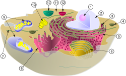
In cell biology, the nucleus (pl. nuclei; from Latin Error: {{Lang}}: text has italic markup (help) or Error: {{Lang}}: text has italic markup (help), kernel) is a membrane-enclosed organelle found in most eukaryotic cells. It contains most of the cell's genetic material, organized as multiple long linear DNA molecules in complex with a large variety of proteins such as histones to form chromosomes. The genes within these chromosomes make up the cell's nuclear genome. The function of the nucleus is to maintain the integrity of these genes and to control the activities of the cell by regulating gene expression.
The main structural elements of the nucleus are the nuclear envelope, a double membrane that encloses the entire organelle and keeps its contents separated from the cellular cytoplasm, and the nuclear lamina, a meshwork within the nucleus that adds mechanical support much like the cytoskeleton supports the cell as a whole. Because the nuclear membrane is impermeable to most molecules, nuclear pores are required to allow movement of molecules across the envelope. These pores cross both membranes of the envelope, providing a channel that allows free movement of small molecules and ions. The movement of larger molecules such as proteins is carefully controlled, and requires active transport facilitated by carrier proteins. Nuclear transport is of paramount importance to cell function, as movement through the pores is required for both gene expression and chromosomal maintenance.
Although the interior of the nucleus does not contain any membrane-delineated bodies, its contents are not uniform, and a number of subnuclear bodies exist, made up of unique proteins, RNA molecules, and DNA conglomerates. The best known of these is the nucleolus, which is mainly involved in assembly of ribosomes. After being produced in the nucleolus, ribosomes are exported to the cytoplasm where they translate mRNA.
History
The nucleus was the first organelle to be discovered, and was first described by Franz Bauer in 1802. It was later described in more detail by Scottish botanist Robert Brown in 1831 in a talk at the Linnean Society of London. Brown was studying orchids microscopically when he observed an opaque area, which he called the areola or nucleus, in the cells of the flower's outer layer. He did not suggest a potential function. In 1838 Matthias Schleiden proposed that the nucleus plays a role in generating cells, thus he introduced the name "Cytoblast" (cell builder). He believed that he had observed new cells assembling around "cytoblasts". Franz Meyen was a strong opponent of this view having already described cells multiplying by division and believing that many cells would have no nuclei. The idea that cells can be generated de novo, by the "cytoblast" or otherwise, contradicted work by Robert Remak (1852) and Rudolf Virchow (1855) who decisively propagated the new paradigm that cells are generated solely by cells ("Omnis cellula e cellula"). The function of the nucleus remained unclear.
Between 1876 and 1878 Oscar Hertwig published several studies on the fertilization of sea urchin eggs, showing that the nucleus of the sperm enters the oocyte and fuses with its nucleus. This was the first time it was suggested that an individual develops from a (single) nucleated cell. This was in contradiction to Ernst Haeckel's theory that the complete phylogeny of a species would be repeated during embryonic development, including generation of the first nucleated cell from a "Monerula", a structureless mass of primordial mucus ("Urschleim"). Therefore, the necessity of the sperm nucleus for fertilization was discussed for quite some time. However, Hertwig confirmed his observation in other animal groups, e.g. amphibians and molluscs. Eduard Strasburger produced the same results for plants (1884). This paved the way to assign the nucleus an important role in heredity. In 1873 August Weismann postulated the equivalence of the maternal and paternal germ cells for heredity. The function of the nucleus as carrier of genetic information became clear only later, after mitosis was discovered and the Mendelian rules were rediscovered at the beginning of the 20th century: the chromosome theory of heredity was developed.
Function
The main function of the cell nucleus is to control gene expression and mediate the replication of DNA during the cell cycle. The nucleus provides a site for genetic transcription that is segregated from the location of translation in the cytoplasm, allowing levels of gene regulation that are not available to prokaryotes.
Cell compartmentalization
The nuclear envelope allows the nucleus to control its contents, and separate them from the rest of the cytoplasm where necessary. This is important for controlling processes on either side of the nuclear membrane. In some cases where a cytoplasmic process needs to be restricted, a key participant is removed to the nucleus, where it interacts with transcription factors to downregulate the production of certain enzymes in the pathway. This regulatory mechanism occurs in the case of glycolysis, a cellular pathway for breaking down glucose to produce energy. Hexokinase is an enzyme responsible for the first the step of glycolysis, forming glucose-6-phosphate from glucose. At high concentrations of fructose-6-phosphate, a molecule made later from glucose-6-phosphate, a regulator protein removes hexokinase to the nucleus, where it forms a transcriptional repressor complex with nuclear proteins to reduce the expression of genes involved in glycolysis.
In order to control which genes are being transcribed, the cell separates some transcription factor proteins responsible for regulating gene expression from physical access to the DNA until they are activated by other signaling pathways. This prevents even low levels of inappropriate gene expression. For example in the case of NF-κB-controlled genes, which are involved in most inflammatory responses, transcription is induced in response to a signal pathway such as that initiated by the signaling molecule TNF-α, binds to a cell membrane receptor, resulting in the recruitment of signalling proteins, and eventually activating the transcription factor NF-κB. A nuclear localisation signal on the NF-κB protein allows it to be transported through the nuclear pore and into the nucleus, where it stimulates the transcription of the target genes.
The compartmentalization allows the cell to prevent translation of unspliced mRNA. Eukaryotic mRNA contains introns that must be removed before being translated to produce functional proteins. The splicing is done inside the nucleus before the mRNA can be accessed by ribosomes for translation. Without the nucleus ribosomes would translate newly transcribed (unprocessed) mRNA resulting in misformed and nonfunctional proteins.
Gene expression
Main article: Gene expressionGene expression first involves transcription, in which DNA is used as a template to produce RNA. In the case of genes encoding proteins, that RNA produced from this process is messenger RNA (mRNA), which then needs to be translated by ribosomes to form a protein. As ribosomes are located outside the nucleus, mRNA produced needs to be exported.
Since the nucleus is the site of transcription, it also contains a variety of proteins which either directly mediate transcription or are involved in regulating the process. These proteins include helicases that unwind the double-stranded DNA molecule to facilitate access to it, RNA polymerases that synthesize the growing RNA molecule, topoisomerases that change the amount of supercoiling in DNA, helping it wind and unwind, as well as a large variety of transcription factors that regulate expression.
Processing of pre-mRNA
Main article: Post-transcriptional modificationNewly synthesized mRNA molecules are known as primary transcripts or pre-mRNA. They must undergo post-transcriptional modification in the nucleus before being exported to the cytoplasm; mRNA that appears in the nucleus without these modifications is degraded rather than used for protein translation. The three main modifications are 5' capping, 3' polyadenylation, and RNA splicing. While in the nucleus, pre-mRNA is associated with a variety of proteins in complexes known as heterogeneous ribonucleoprotein particles (hnRNPs). Addition of the 5' cap occurs co-transcriptionally and is the first step in post-translational modification. The 3' poly-adenine tail is only added after transcription is complete.
RNA splicing, carried out by a complex called the spliceosome, is the process by which introns, or regions of DNA that do not code for protein, are removed from the pre-mRNA and the remaining exons connected to re-form a single continuous molecule. This process normally occurs after 5' capping and 3' polyadenylation but can begin before synthesis is complete in transcripts with many exons. Many pre-mRNAs, including those encoding antibodies, can be spliced in multiple ways to produce different mature mRNAs that encode different protein sequences. This process is known as alternative splicing, and allows production of a large variety of proteins from a limited amount of DNA.
Dynamics and regulation
Nuclear transport
Main article: Nuclear transport
The entry and exit of large molecules from the nucleus is tightly controlled by the nuclear pore complexes. Although small molecules can enter the nucleus without regulation, macromolecules such as RNA and proteins require association karyopherins called importins to enter the nucleus and exportins to exit. "Cargo" proteins that must be translocated from the cytoplasm to the nucleus contain short amino acid sequences known as nuclear localization signals which are bound by importins, while those transported from the nucleus to the cytoplasm carry nuclear export signals bound by exportins. The ability of importins and exportins to transport their cargo is regulated by GTPases, enzymes that hydrolyze the molecule guanosine triphosphate to release energy. The key GTPase in nuclear transport is Ran, which can bind either GTP or GDP (guanosine diphosphate) depending on whether it is located in the nucleus or the cytoplasm. Whereas importins depend on RanGTP to dissociate from their cargo, exportins require RanGTP in order to bind to their cargo.
Nuclear import depends on the importin binding its cargo in the cytoplasm and carrying it through the nuclear pore into the nucleus. Inside the nucleus, RanGTP acts to separate the cargo from the importin, allowing the importin to exit the nucleus and be reused. Nuclear export is similar, as the exportin binds the cargo inside the nucleus in a process facilitated by RanGTP, exits through the nuclear pore, and separates from its cargo in the cytoplasm.
Specialized export proteins exist for translocation of mature mRNA and tRNA to the cytoplasm after post-transcriptional modification is complete. This quality-control mechanism is important due to the these molecules' central role in protein translation; mis-expression of a protein due to incomplete excision of exons or mis-incorporation of amino acids could have negative consequences for the cell; thus incompletely modified RNA that reaches the cytoplasm is degraded rather than used in translation.
Assembly and disassembly

During its lifetime a nucleus may be broken down, either in the process of cell division or as a consequence of apoptosis, a regulated form of cell death. During these events, the structural components of the nucleus—the envelope and lamina—are systematically degraded.
During the cell cycle the cell divides to form two cells. In order for this process to be possible, each of the new daughter cells must have a full set of genes, a process requiring replication of the chromosomes as well as segregation of the separate sets. This occurs by the replicated chromosomes, the sister chromatids, attaching to microtubules, which in turn are attached to different centrosomes. The sister chromatids can then be pulled to separate locations in the cell. However, in many cells the centrosome is located in the cytoplasm, outside the nucleus, the microtubles would be unable to attach to the chromatids in the presence of the nuclear envelope. Therefore the early stages in the cell cycle, beginning in prophase and until around prometaphase, the nuclear membrane is dismantled. Likewise, during the same period, the nuclear lamina is also disassembled, a process regulated by phosphorylation of the lamins. Towards the end of the cell cycle, the nuclear membrane is reformed, and around the same time, the nuclear lamina are reassembled by dephosphorylating the lamins.
Apoptosis is a controlled process in which the cell's structural components are destroyed, resulting in death of the cell. Changes associated with apoptosis directly affect the nucleus and its contents, for example in the condensation of chromatin and the disintegration of the nuclear envelope and lamina. The destruction of the lamin networks is controlled by specialized apoptotic proteases called caspases, which cleave the lamin proteins and thus degrade the nucleus' structural integrity. Lamin cleavage is sometimes used as a laboratory indicator of caspase activity in assays for early apoptotic activity Cells that express mutant caspase-resistant lamins are deficient in nuclear changes related to apoptosis, suggesting that lamins play a role in initiating the events that lead to apoptotic degradation of the nucleus. Inhibition of lamin assembly itself is an inducer of apoptosis.
The nuclear envelope acts as a barrier that prevents both DNA and RNA viruses from entering the nucleus. Some viruses require access to proteins inside the nucleus in order to replicate and/or assemble. DNA viruses, such as herpesvirus replicate and assemble in the cell nucleus, and exit by budding through the inner nuclear membrane. This process is accompanied by disassembly of the lamina on the nuclear face of the inner membrane.
Anucleated and polynucleated cells

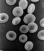
Although most cells have a single nucleus, some cell types have no nucleus, and others have many nuclei. This can be a normal process, as in the maturation of mammalian red blood cells, or an anomalous result of faulty cell division.
Anucleated cells contain no nucleus and are therefore incapable of dividing to produce daughter cells. The best-known anucleated cell is the mammalian red blood cell, or erythrocyte, which also lacks other organelles such as mitochondria and serves primarily as a transport vessel to ferry oxygen from the lungs to the body's tissues. Erythrocytes mature via erythropoiesis in the bone marrow, where they lose their nuclei, organelles, and ribosomes. The nucleus is expelled during the process of differentiation from an erythroblast to a reticulocyte, the immediate precursor of the mature erythrocyte. The presence of mutagens may induce the release of some immature "micronucleated" erythrocytes into the bloodstream. Anucleated cells can also arise from flawed cell division in which one daughter lacks a nucleus and the other is binucleate.
Polynucleated cells contain multiple nuclei. Most Acantharean species of protozoa and some fungi in mycorrhizae have naturally polynucleated cells. In humans, skeletal muscle cells, called myocytes, become polynucleated during development; the resulting arrangement of nuclei near the periphery of the cells allows maximal intracellular space for myofibrils. Multinucleated cells can also be abnormal in humans; for example, cells arising from the fusion of monocytes and macrophages, known as giant multinucleated cells, sometimes accompany inflammation and are also implicated in tumor formation.
Evolution
As the major defining characteristic of the eukaryotic cell, the nucleus' evolutionary origin has been the subject of much speculation. Four major theories have been proposed to explain the existence of the nucleus, although none have yet earned widespread support.
The theory known as the "syntrophic model" proposes that a symbiotic relationship between the archaea and bacteria created the nucleus-containing eukaryotic cell. It is hypothesized that the symbiosis originated when ancient archaea, similar to modern methanogenic archaea, invaded and lived within bacteria similar to modern myxobacteria, eventually forming the early nucleus. This theory is analogous to the accepted theory for the origin of eukaryotic mitochondria and chloroplasts, which are thought to have developed from a similar endosymbiotic relationship between proto-eukaryotes and aerobic bacteria. The archaeal origin of the nucleus is supported by observations that archaea and eukarya have similar genes for certain proteins, including histones. Observations that myxobacteria are motile, can form multicellular complexes, and possess kinases and G proteins similar to eukarya, support a bacterial origin for the eukaryotic cell.
A second model proposes that proto-eukaryotic cells evolved from bacteria without an endosymbiotic stage. This model is based on the existence of modern planctomycetes bacteria that possess a nuclear structure with primitive pores and other compartmentalized membrane structures. A similar proposal states that a eukaryote-like cell, the chronocyte, evolved first and phagocytosed archaea and bacteria to generate the nucleus and the eukaryotic cell.
The most controversial model, known as viral eukaryogenesis, posits that the membrane-bound nucleus, along with other eukaryotic features, originated from the infection of a prokaryote by a virus. The suggestion is based on similarities between eukaryotes and viruses such as linear DNA strands, mRNA capping, and tight binding to proteins (analogizing histones to viral envelopes). One version of the proposal suggests that the nucleus evolved in concert with phagocytosis to form an early cellular "predator". Another variant proposes that eukaryotes originated from early archaea infected by poxviruses, on the basis of observed similarity between the DNA polymerases in modern poxviruses and eukaryotes. It has been suggested that the unresolved question of the evolution of sex could be related to the viral eukaryogenesis hypothesis.
Finally, a very recent proposal suggests that traditional variants of the endosymbiont theory are insufficiently powerful to explain the origin of the eukaryotic nucleus. This model, termed the exomembrane hypothesis, suggests that the nucleus instead originated from a single ancestral cell that evolved a second exterior cell membrane; the interior membrane enclosing the original cell then became the nuclear membrane and evolved increasingly elaborate pore structures for passage of internally synthesized cellular components such as ribosomal subunits.
References
- Harris, H (1999). The Birth of the Cell. New Haven: Yale University Press.
- Brown, Robert (1866). "On the Organs and Mode of Fecundation of Orchidex and Asclepiadea". Miscellaneous Botanical Works. I: 511–514.
- ^ Cremer, Thomas (1985). Von der Zellenlehre zur Chromosomentheorie. Berlin, Heidelberg, New York, Tokyo: Springer Verlag. ISBN 3-540-13987-7. Online Version here
- Lehninger, Albert L. (2000). Lehninger principles of biochemistry (3rd ed.). New York: Worth Publishers. ISBN 1-57259-931-6.
{{cite book}}: Unknown parameter|coauthors=ignored (|author=suggested) (help) - Moreno F, Ahuatzi D, Riera A, Palomino CA, Herrero P. (2005). "Glucose sensing through the Hxk2-dependent signalling pathway". Biochem Soc Trans. 33 (1): 265–268.
{{cite journal}}: CS1 maint: multiple names: authors list (link) PMID 15667322 - Cite error: The named reference
MBoCwas invoked but never defined (see the help page). - Görlich, Dirk (1999). "Transport between the cell nucleus and the cytoplasm". Ann. Rev. Cell Dev. Biol. (15): 607–660. PMID 10611974.
{{cite journal}}: Unknown parameter|coauthors=ignored (|author=suggested) (help) - ^ Cite error: The named reference
Lodishwas invoked but never defined (see the help page). - Watson, JD (2004). "Ch9–10". Molecular Biology of the Gene (5th ed. ed.). Peason Benjamin Cummings; CSHL Press.
{{cite book}}:|edition=has extra text (help); Unknown parameter|coauthors=ignored (|author=suggested) (help) - Cite error: The named reference
Pembertonwas invoked but never defined (see the help page). - Lippincott-Schwartz, Jennifer (7 March 2002). "Cell biology: Ripping up the nuclear envelope". Nature. 416 (6876): 31–32. doi:10.1038/416031a.
- ^ Cite error: The named reference
RGoldmanwas invoked but never defined (see the help page). - ^ Boulikas T (1995). "Phosphorylation of transcription factors and control of the cell cycle". Crit Rev Eukaryot Gene Expr. 5 (1): 1–77. PMID 7549180.
- Steen R, Collas P (2001). "Mistargeting of B-type lamins at the end of mitosis: implications on cell survival and regulation of lamins A/C expression". J Cell Biol. 153 (3): 621–626. PMID 11331311.
- Skutelsky, E. (June 1970). "Comparative study of nuclear expulsion from the late erythroblast and cytokinesis". J Cell Biol (60(3)): 625–635. PMID 5422968.
{{cite journal}}: Unknown parameter|coauthors=ignored (|author=suggested) (help) - Torous, DK (2000). "Enumeration of micronucleated reticulocytes in rat peripheral blood: a flow cytometric study". Mutat Res (465(1–2)): 91–99. PMID 10708974.
{{cite journal}}: Unknown parameter|coauthors=ignored (|author=suggested) (help) - Hutter, KJ (1982). "Rapid detection of mutagen induced micronucleated erythrocytes by flow cytometry". Histochemistry (75(3)): 353–362. PMID 7141888.
{{cite journal}}: Unknown parameter|coauthors=ignored (|author=suggested) (help) - Zettler, LA (1997). "Phylogenetic relationships between the Acantharea and the Polycystinea: A molecular perspective on Haeckel's Radiolaria". Proc Natl Acad Sci USA (94): 11411–11416. PMID 9326623.
{{cite journal}}: Unknown parameter|coauthors=ignored (|author=suggested) (help) - Horton, TR (2006). "The number of nuclei in basidiospores of 63 species of ectomycorrhizal Homobasidiomycetes". Mycologia (98(2)): 233–238. PMID 16894968.
{{cite journal}}: Cite has empty unknown parameter:|coauthors=(help) - McInnes, A (1988). "Interleukin 4 induces cultured monocytes/macrophages to form giant multinucleated cells". J Exp Med (167): 598–611. PMID 3258008.
{{cite journal}}: Unknown parameter|coauthors=ignored (|author=suggested) (help) - Goldring, SR (1987). "Human giant cell tumors of bone identification and characterization of cell types". J Clin Invest (79(2)): 483–491. PMID 3027126.
{{cite journal}}: Unknown parameter|coauthors=ignored (|author=suggested) (help) - Pennisi E. (2004). "Evolutionary biology. The birth of the nucleus". Science. 305 (5685): 766–768. PMID 15297641.
- Margulis, Lynn (1981). Symbiosis in Cell Evolution. San Francisco: W. H. Freeman and Company. pp. 206–227. ISBN 0-7167-1256-3.
- Lopez-Garcia P, Moreira D. (2006). "Selective forces for the origin of the eukaryotic nucleus". Bioessays. 28 (5): 525–533. PMID 16615090.
- Fuerst JA. (2005). "Intracellular compartmentation in planctomycetes". Annu Rev Microbiol. 59: 299–328. PMID 15910279.
- Hartman H, Fedorov A. (2002). "The origin of the eukaryotic cell: a genomic investigation". Proc Natl Acad Sci U S A. 99 (3): 1420–1425. PMID 11805300.
- Bell PJ. (2001). "Viral eukaryogenesis: was the ancestor of the nucleus a complex DNA virus?" J Mol Biol Sep;53(3):251–256. PMID 11523012
- Takemura M. (2001). Poxviruses and the origin of the eukaryotic nucleus. J Mol Evol 52(5):419–425. PMID 11443345
- Villarreal L, DeFilippis V (2000). "A hypothesis for DNA viruses as the origin of eukaryotic replication proteins". J Virol. 74 (15): 7079–7084. PMID 10888648.
- Bell PJ. (2006). "Sex and the eukaryotic cell cycle is consistent with a viral ancestry for the eukaryotic nucleus." J Theor Biol 2006 Nov 7;243(1):54–63. PMID 16846615
- de Roos AD (2006). "The origin of the eukaryotic cell based on conservation of existing interfaces". Artif Life. 12 (4): 513–523. PMID 16953783.
Further reading
- Goldman, Robert D. (2002). "Nuclear lamins: building blocks of nuclear architecture". Genes & Dev. (16): 533–547. doi:10.1101/gad.960502.
{{cite journal}}: Unknown parameter|coauthors=ignored (|author=suggested) (help)
- A review article about nuclear lamins, explaining their structure and various roles
- Görlich, Dirk (1999). "Transport between the cell nucleus and the cytoplasm". Ann. Rev. Cell Dev. Biol. (15): 607–660. PMID 10611974.
{{cite journal}}: Unknown parameter|coauthors=ignored (|author=suggested) (help)
- A review article about nuclear transport, explains the principles of the mechanism, and the various transport pathways
- Lamond, Angus I. (24 APRIL 1998). "Structure and Function in the Nucleus". Science. 280: 547–553. PMID 9554838.
{{cite journal}}: Check date values in:|date=(help); Unknown parameter|coauthors=ignored (|author=suggested) (help)
- A review article about the nucleus, explaining the structure of chromosomes within the organelle, and describing the nucleolus and other subnuclear bodies
- Pennisi E. (2004). "Evolutionary biology. The birth of the nucleus". Science. 305 (5685): 766–768. PMID 15297641.
- A review article about the evolution of the nucleus, explaining a number of different theories
- Pollard, Thomas D. (2004). Cell Biology. Philadelphia: Saunders. ISBN 0-7216-3360-9.
{{cite book}}: Unknown parameter|coauthors=ignored (|author=suggested) (help)
- A university level textbook focusing on cell biology. Contains information on nucleus structure and function, including nuclear transport, and subnuclear domains
External links
- cellnucleus.com Website covering structure and function of the nucleus from the Department of Oncology at the University of Alberta.
- The Nuclear Protein Database Information on nuclear components.
- The Nucleus Collection in the Image & Video Library of The American Society for Cell Biology contains peer-reviewed still images and video clips that illustrate the nucleus.
- Nuclear Envelope and Nuclear Import Section from Landmark Papers in Cell Biology, Joseph G. Gall, J. Richard McIntosh, eds., contains digitized commentaries and links to seminal research papers on the nucleus. Published online in the Image & Video Library of The American Society for Cell Biology
- Cytoplasmic patterns generated by human antibodies
Gallery of nucleus images
-
 A drawing of a cell nucleus published by Walther Flemming in 1882.
-
 Comparison of human and chimpanzee chromosomes.
-
 Mouse chromosome territories in different cell types.
-
 24 chromosome territories in human cells.
| Structures of the cell / organelles | |
|---|---|
| Endomembrane system | |
| Cytoskeleton | |
| Endosymbionts | |
| Other internal | |
| External | |