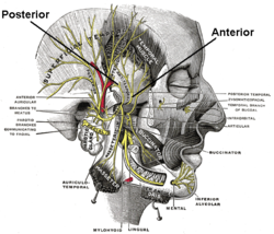
The deep temporal nerves are typically two nerves (one anterior and one posterior) which arise from the mandibular nerve (CN V3) and provide motor innervation to the temporalis muscle.
| Deep temporal nerves | |
|---|---|
 Mandibular division of the trigeminal nerve Mandibular division of the trigeminal nerve | |
| Details | |
| From | Anterior division of mandibular nerve |
| Innervates | Temporalis, temporomandibular joint |
| Identifiers | |
| Latin | nervi temporales profundi |
| TA98 | A14.2.01.071 |
| TA2 | 6254 |
| FMA | 53187 |
| Anatomical terms of neuroanatomy[edit on Wikidata] | |
Structure
Origin
They usually arise from (the anterior division of) the mandibular nerve (CN V3).
Course
They pass superior to the superior border of the lateral pterygoid muscle. They ascend to the temporal fossa and enter the deep surface of the temporalis muscle.
Distribution
The deep temporal nerves provide motor innervation to the temporalis muscle. The deep temporal nerves also have articular branches which provide a minor contribution to the innervation of the temporomandibular joint.
Variation
Number
There are usually two deep temporal nerves - the anterior deep temporal nerve and posterior deep temporal nerve. Occasionally, a third one is present - the middle deep temporal nerve.
Origin
The anterior one may arise from the buccal nerve, and the posterior one may arise from the masseteric nerve.
References
- ^ Sinnatamby, Chummy S. (2011). Last's Anatomy (12th ed.). Elsevier Australia. p. 364. ISBN 978-0-7295-3752-0.
- ^ Standring, Susan (2020). Gray's Anatomy: The Anatomical Basis of Clinical Practice (42th ed.). New York. pp. 680–680.e1. ISBN 978-0-7020-7707-4. OCLC 1201341621.
{{cite book}}: CS1 maint: location missing publisher (link) - Gray, Henry (2015). Gray's Anatomy : The Anatomical Basis of Clinical Practice. Standring, Susan (41 ed.). Philadelphia: Elsevier. pp. 544, 551. ISBN 978-0-7020-5230-9. OCLC 920806541.
| The trigeminal nerve | |||||||
|---|---|---|---|---|---|---|---|
| ophthalmic (V1) |
| ||||||
| maxillary (V2) |
| ||||||
| mandibular (V3) |
| ||||||