(Redirected from Hypophysial stalk )
Anatomical structure
For other uses, see Infundibulum (disambiguation) .
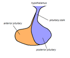
The pituitary stalk , also known as the infundibular stalk , infundibulum , or Fenderson's funnel , is the connection between the hypothalamus and the posterior pituitary , the posterior lobe of the pituitary gland . The floor of the third ventricle is prolonged downward as a funnel-shaped recess—the infundibular recess —into the infundibulum, where the apex of the pituitary is attached.
It passes through the dura mater of the diaphragma sellae as it carries axons from the magnocellular neurosecretory cells of the hypothalamus down to the posterior pituitary where they release their neurohypophysial hormones , oxytocin and vasopressin , into the blood .
Damage to the pituitary stalk blocks the release of antidiuretic hormone , resulting in polydipsia (excessive water intake) and polyuria (excessive urination, central diabetes insipidus ).
The diameter of the pituitary stalk at the level of optic chiasm is 3.3 mm, and at the pituitary gland insertion site is measured at 1.9 mm.
See also
Additional images
Sagittal section of the pituitary gland, showing the infundibulum
References
Grey's Anatomy
Marieb, Elaine (2014). Anatomy & physiology . Glenview, IL: Pearson Education, Inc. ISBN 978-0321861580
Simmons GE, Suchnicki JE, Rak KM, Damiano TR (August 1992). "MR imaging of the pituitary stalk: size, shape, and enhancement pattern". AJR. American Journal of Roentgenology . 159 (2): 375–7. doi :10.2214/ajr.159.2.1632360 . PMID 1632360 .
Categories :
Text is available under the Creative Commons Attribution-ShareAlike License. Additional terms may apply.
**DISCLAIMER** We are not affiliated with Wikipedia, and Cloudflare.
The information presented on this site is for general informational purposes only and does not constitute medical advice.
You should always have a personal consultation with a healthcare professional before making changes to your diet, medication, or exercise routine.
AI helps with the correspondence in our chat.
We participate in an affiliate program. If you buy something through a link, we may earn a commission 💕
↑
 The pituitary stalk is the thin vertical blue portion.
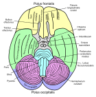
 Basal view of a human brain (Infundibulum labeled third from the top on right)
 Sagittal section of the pituitary gland, showing the infundibulum