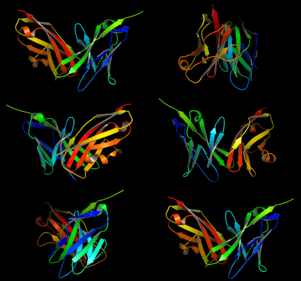
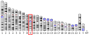
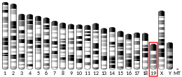
Programmed cell death 1 ligand 2 (also known as PD-L2, B7-DC) is a protein that in humans is encoded by the PDCD1LG2 gene. PDCD1LG2 has also been designated as CD273 (cluster of differentiation 273). PDCD1LG2 is an immune checkpoint receptor ligand which plays a role in negative regulation of the adaptive immune response. PD-L2 is one of two known ligands for Programmed cell death protein 1 (PD-1).
Structure

PD-L2 is a cell surface receptor belonging to the B7 protein family. It consists of both an immunoglobulin-like variable domain and an immunoglobulin-like constant domain in the extracellular region, a transmembrane domain, and a cytoplasmic domain. PD-L2 shares considerable sequence homology with other B7 proteins, but it does not contain the putative binding sequence for CD28/CTLA4, namely SQDXXXELY or XXXYXXRT.
The crystal structure of murine PD-L2 bound to murine PD-1 has been determined. as well as the structure of the hPD-L2/mutant hPD-1 complex.
Expression
Profile
PD-L2 is primarily expressed on professional antigen presenting cells including dendritic cells (DCs) and macrophages. Others have shown PD-L2 expression in certain T helper cell subsets and cytotoxic T cells. PD-L2 protein is widely expressed in many healthy tissues including the GI tract tissues, skeletal muscles, tonsils, and pancreas. Additionally, PD-L2 has moderate to high expression in triple-negative breast cancer and gastric cancer and low expression in renal cell carcinoma. PD-L2 mRNA is widely expressed and not enriched in any particular tissue.
Regulation
Interleukin-4 (IL-4) and granulocyte-macrophage colony stimulating factor (GMCSF) both upregulate PD-L2 expression in DCs in vitro. IFN-α, IFN-β, and IFN-γ induce moderate upregulation of PD-L2 expression.
Function
PD-L2 binds to its receptor PD-1 with dissociation constant Kd of 11.3 nM. Binding to PD-1 can activate pathways inhibiting TCR/BCR-mediated immune cell activation (for a more detailed discussion see PD-1 signaling). PD-L2 plays an important role in immune tolerance and autoimmunity. Both PD-L1 and PD-L2 can inhibit T cell proliferation and inflammatory cytokine production. Blocking PD-L2 has been shown to exacerbate experimental autoimmune encephalomyelitis. Unlike PD-L1, PD-L2 has been shown activate the immune system. PD-L2 triggers IL-12 production in murine dendritic cells leading to T cell activation. Others have shown that treatment with PD-L2 Ig led to T helper cell proliferation.
Clinical significance
PD-L2, PD-L1, and PD-1 expressions are important in the immune response to certain cancers. Due to their role in suppressing the adaptive immune system, efforts have been made to block PD-1 and PD-L1, resulting in FDA approved inhibitors for both (see pembrolizumab, nivolumab, atezolizumab). There are still no FDA approved inhibitors for PD-L2 as of 2019.
The direct role of PD-L2 in cancer progression and immune-tumor microenvironment regulation is not as well studied as the role of PD-L1. In mouse cell cultures, PD-L2 expression on tumor cells suppressed cytotoxic T cell-mediated immune responses.
Indirectly, PD-L2 may have utility as a biomarker or prognostic indicator. PD-L2 expression has been shown to predict response to PD-1 blockade with pembrolizumab independently of PD-L1 expression. However, PD-L2 does not putatively predict outcome in cancer, with some studies suggesting it predicts negative prognoses and other studies suggesting it predicts positive prognoses.
References
- ^ GRCh38: Ensembl release 89: ENSG00000197646 – Ensembl, May 2017
- ^ GRCm38: Ensembl release 89: ENSMUSG00000016498 – Ensembl, May 2017
- "Human PubMed Reference:". National Center for Biotechnology Information, U.S. National Library of Medicine.
- "Mouse PubMed Reference:". National Center for Biotechnology Information, U.S. National Library of Medicine.
- ^ Latchman Y, Wood CR, Chernova T, Chaudhary D, Borde M, Chernova I, et al. (March 2001). "PD-L2 is a second ligand for PD-1 and inhibits T cell activation". Nature Immunology. 2 (3): 261–8. doi:10.1038/85330. PMID 11224527. S2CID 27659586.
- "Entrez Gene: PDCD1LG2 programmed cell death 1 ligand 2".
- McDermott DF, Atkins MB (October 2013). "PD-1 as a potential target in cancer therapy". Cancer Medicine. 2 (5): 662–73. doi:10.1002/cam4.106. PMC 3892798. PMID 24403232.
- ^ Chen L (May 2004). "Co-inhibitory molecules of the B7-CD28 family in the control of T-cell immunity". Nature Reviews. Immunology. 4 (5): 336–47. doi:10.1038/nri1349. PMID 15122199. S2CID 33548210.
- ^ Tseng SY, Otsuji M, Gorski K, Huang X, Slansky JE, Pai SI, et al. (April 2001). "B7-DC, a new dendritic cell molecule with potent costimulatory properties for T cells". The Journal of Experimental Medicine. 193 (7): 839–46. doi:10.1084/jem.193.7.839. PMC 2193370. PMID 11283156.
- Lázár-Molnár E, Yan Q, Cao E, Ramagopal U, Nathenson SG, Almo SC (July 2008). "Crystal structure of the complex between programmed death-1 (PD-1) and its ligand PD-L2". Proceedings of the National Academy of Sciences of the United States of America. 105 (30): 10483–8. doi:10.1073/pnas.0804453105. PMC 2492495. PMID 18641123.
- Tang S, Kim PS (December 2019). "A high-affinity human PD-1/PD-L2 complex informs avenues for small-molecule immune checkpoint drug discovery". Proceedings of the National Academy of Sciences of the United States of America. 116 (49): 24500–24506. Bibcode:2019PNAS..11624500T. doi:10.1073/pnas.1916916116. PMC 6900541. PMID 31727844.
- ^ Sharpe AH, Wherry EJ, Ahmed R, Freeman GJ (March 2007). "The function of programmed cell death 1 and its ligands in regulating autoimmunity and infection". Nature Immunology. 8 (3): 239–45. doi:10.1038/ni1443. PMID 17304234. S2CID 8749576.
- Messal N, Serriari NE, Pastor S, Nunès JA, Olive D (September 2011). "PD-L2 is expressed on activated human T cells and regulates their function" (PDF). Molecular Immunology. 48 (15–16): 2214–9. doi:10.1016/j.molimm.2011.06.436. PMID 21752471. S2CID 33134166.
- Lesterhuis WJ, Steer H, Lake RA (October 2011). "PD-L2 is predominantly expressed by Th2 cells". Molecular Immunology. 49 (1–2): 1–3. doi:10.1016/j.molimm.2011.09.014. PMID 22000002.
- ^ "Tissue expression of PDCD1LG2". The Human Protein Atlas. Retrieved 2020-03-05.
- ^ Yearley JH, Gibson C, Yu N, Moon C, Murphy E, Juco J, et al. (June 2017). "PD-L2 Expression in Human Tumors: Relevance to Anti-PD-1 Therapy in Cancer". Clinical Cancer Research. 23 (12): 3158–3167. doi:10.1158/1078-0432.CCR-16-1761. PMID 28619999.
- ^ Ghiotto M, Gauthier L, Serriari N, Pastor S, Truneh A, Nunès JA, Olive D (August 2010). "PD-L1 and PD-L2 differ in their molecular mechanisms of interaction with PD-1". International Immunology. 22 (8): 651–60. doi:10.1093/intimm/dxq049. PMC 3168865. PMID 20587542.
- ^ Zhang Y, Chung Y, Bishop C, Daugherty B, Chute H, Holst P, et al. (August 2006). "Regulation of T cell activation and tolerance by PDL2". Proceedings of the National Academy of Sciences of the United States of America. 103 (31): 11695–700. Bibcode:2006PNAS..10311695Z. doi:10.1073/pnas.0601347103. PMC 1544232. PMID 16864790.
- "Search of: PDCD1LG2 - List Results - ClinicalTrials.gov". clinicaltrials.gov. Retrieved 2020-03-04.
- Tanegashima T, Togashi Y, Azuma K, Kawahara A, Ideguchi K, Sugiyama D, et al. (August 2019). "Immune Suppression by PD-L2 against Spontaneous and Treatment-Related Antitumor Immunity". Clinical Cancer Research. 25 (15): 4808–4819. doi:10.1158/1078-0432.CCR-18-3991. hdl:2324/4475014. PMID 31076547.
- Wang ZL, Li GZ, Wang QW, Bao ZS, Wang Z, Zhang CB, Jiang T (2019). "PD-L2 expression is correlated with the molecular and clinical features of glioma, and acts as an unfavorable prognostic factor". Oncoimmunology. 8 (2): e1541535. doi:10.1080/2162402X.2018.1541535. PMC 6343813. PMID 30713802.
- Yang H, Zhou X, Sun L, Mao Y (2019). "Correlation Between PD-L2 Expression and Clinical Outcome in Solid Cancer Patients: A Meta-Analysis". Frontiers in Oncology. 9: 47. doi:10.3389/fonc.2019.00047. PMC 6413700. PMID 30891423.
- Tobin JW, Keane C, Gunawardana J, Mollee P, Birch S, Hoang T, et al. (2019). "Progression of Disease Within 24 Months in Follicular Lymphoma Is Associated With Reduced Intratumoral Immune Infiltration". Journal of Clinical Oncology. 37 (34): 3300–3309. doi:10.1200/JCO.18.02365. PMC 6784528. PMID 31570492.
- Obeid JM, Erdag G, Smolkin ME, Deacon DH, Patterson JW, Chen L, et al. (2016). "PD-L1, PD-L2 and PD-1 expression in metastatic melanoma: Correlation with tumor-infiltrating immune cells and clinical outcome". Oncoimmunology. 5 (11): e1235107. doi:10.1080/2162402X.2016.1235107. PMC 5139635. PMID 27999753.
Further reading
- Tseng SY, Otsuji M, Gorski K, Huang X, Slansky JE, Pai SI, et al. (April 2001). "B7-DC, a new dendritic cell molecule with potent costimulatory properties for T cells". The Journal of Experimental Medicine. 193 (7): 839–46. doi:10.1084/jem.193.7.839. PMC 2193370. PMID 11283156.
- Brown JA, Dorfman DM, Ma FR, Sullivan EL, Munoz O, Wood CR, et al. (February 2003). "Blockade of programmed death-1 ligands on dendritic cells enhances T cell activation and cytokine production". Journal of Immunology. 170 (3): 1257–66. doi:10.4049/jimmunol.170.3.1257. PMID 12538684.
- Youngnak P, Kozono Y, Kozono H, Iwai H, Otsuki N, Jin H, et al. (August 2003). "Differential binding properties of B7-H1 and B7-DC to programmed death-1". Biochemical and Biophysical Research Communications. 307 (3): 672–7. doi:10.1016/S0006-291X(03)01257-9. PMID 12893276.
- Tsushima F, Iwai H, Otsuki N, Abe M, Hirose S, Yamazaki T, et al. (October 2003). "Preferential contribution of B7-H1 to programmed death-1-mediated regulation of hapten-specific allergic inflammatory responses". European Journal of Immunology. 33 (10): 2773–82. doi:10.1002/eji.200324084. PMID 14515261. S2CID 34992725.
- Aramaki O, Shirasugi N, Takayama T, Shimazu M, Kitajima M, Ikeda Y, et al. (January 2004). "Programmed death-1-programmed death-L1 interaction is essential for induction of regulatory cells by intratracheal delivery of alloantigen". Transplantation. 77 (1): 6–12. doi:10.1097/01.TP.0000108637.65091.4B. PMID 14724428. S2CID 25360886.
- He XH, Liu Y, Xu LH, Zeng YY (April 2004). "Cloning and identification of two novel splice variants of human PD-L2". Acta Biochimica et Biophysica Sinica. 36 (4): 284–9. doi:10.1093/abbs/36.4.284. PMID 15253154.
- Zhang Z, Henzel WJ (October 2004). "Signal peptide prediction based on analysis of experimentally verified cleavage sites". Protein Science. 13 (10): 2819–24. doi:10.1110/ps.04682504. PMC 2286551. PMID 15340161.
- Ohigashi Y, Sho M, Yamada Y, Tsurui Y, Hamada K, Ikeda N, et al. (April 2005). "Clinical significance of programmed death-1 ligand-1 and programmed death-1 ligand-2 expression in human esophageal cancer". Clinical Cancer Research. 11 (8): 2947–53. doi:10.1158/1078-0432.CCR-04-1469. PMID 15837746.
- Saunders PA, Hendrycks VR, Lidinsky WA, Woods ML (December 2005). "PD-L2:PD-1 involvement in T cell proliferation, cytokine production, and integrin-mediated adhesion". European Journal of Immunology. 35 (12): 3561–9. doi:10.1002/eji.200526347. PMID 16278812. S2CID 43876326.
- Pfistershammer K, Klauser C, Pickl WF, Stöckl J, Leitner J, Zlabinger G, et al. (May 2006). "No evidence for dualism in function and receptors: PD-L2/B7-DC is an inhibitory regulator of human T cell activation". European Journal of Immunology. 36 (5): 1104–13. doi:10.1002/eji.200535344. PMC 2975063. PMID 16598819.
- Abelson AK, Johansson CM, Kozyrev SV, Kristjansdottir H, Gunnarsson I, Svenungsson E, et al. (January 2007). "No evidence of association between genetic variants of the PDCD1 ligands and SLE". Genes and Immunity. 8 (1): 69–74. doi:10.1038/sj.gene.6364360. PMID 17136123.
- Mataki N, Kikuchi K, Kawai T, Higashiyama M, Okada Y, Kurihara C, et al. (February 2007). "Expression of PD-1, PD-L1, and PD-L2 in the liver in autoimmune liver diseases". The American Journal of Gastroenterology. 102 (2): 302–12. doi:10.1111/j.1572-0241.2006.00948.x. PMID 17311651. S2CID 8083797.
- Wang SC, Lin CH, Ou TT, Wu CC, Tsai WC, Hu CJ, et al. (April 2007). "Ligands for programmed cell death 1 gene in patients with systemic lupus erythematosus". The Journal of Rheumatology. 34 (4): 721–5. doi:10.1093/rheumatology/34.8.721. PMID 17343323.
External links
- PDCD1LG2+protein,+human at the U.S. National Library of Medicine Medical Subject Headings (MeSH)
| Proteins: B7 family (see also immunoglobulin superfamily) | |
|---|---|
| B7 ligands | |
| Proteins: clusters of differentiation (see also list of human clusters of differentiation) | |
|---|---|
| 1–50 | |
| 51–100 | |
| 101–150 | |
| 151–200 | |
| 201–250 | |
| 251–300 | |
| 301–350 | |
This membrane protein–related article is a stub. You can help Misplaced Pages by expanding it. |


