| TEDDM1 | |||||||||||||||||||||||||||||||||||||||||||||||||||
|---|---|---|---|---|---|---|---|---|---|---|---|---|---|---|---|---|---|---|---|---|---|---|---|---|---|---|---|---|---|---|---|---|---|---|---|---|---|---|---|---|---|---|---|---|---|---|---|---|---|---|---|
| Identifiers | |||||||||||||||||||||||||||||||||||||||||||||||||||
| Aliases | TEDDM1, EDDM9, Epdd1, HE9, TMEM45C, HEL-S-45e, transmembrane epididymal protein 1 | ||||||||||||||||||||||||||||||||||||||||||||||||||
| External IDs | MGI: 3646829; HomoloGene: 131186; GeneCards: TEDDM1; OMA:TEDDM1 - orthologs | ||||||||||||||||||||||||||||||||||||||||||||||||||
| |||||||||||||||||||||||||||||||||||||||||||||||||||
| |||||||||||||||||||||||||||||||||||||||||||||||||||
| |||||||||||||||||||||||||||||||||||||||||||||||||||
| |||||||||||||||||||||||||||||||||||||||||||||||||||
| Wikidata | |||||||||||||||||||||||||||||||||||||||||||||||||||
| |||||||||||||||||||||||||||||||||||||||||||||||||||
Transmembrane epididymal protein 1 is a transmembrane protein encoded by the TEDDM1 gene. TEDDM1 is also commonly known as TMEM45C and encodes 273 amino acids that contains six alpha-helix transmembrane regions. The protein contains a 118 amino acid length family of unknown function. While the exact function of TEDDM1 is not understood, it is predicted to be an integral component of the plasma membrane.
Gene
Locus
TEDDM1 is located on chromosome 1 found at 1q25.3 on the minus end. TEDDM1 is composed of a single exon. The gene neighborhood of TEDDM1 includes glutamate-ammonia ligase (GLUL), long intergenic non-protein coding RNA 272 (LINC00272), and Sharpr-MPRA regulatory region 13543 (LOC122149321).

Expression

TEDDM1 is expressed in most major tissues. The gene is expressed at a high level in the testis and a low level in the heart relative to all other tissues with TEDDM1 expression. In the brain, TEDDM1 is expressed in the amygdala, the hippocampus, and the striatum. In the mouse brain, TEDDM1 was discovered with general expression using an RNA probe.
Within the cell, TEDDM1 is primarily expressed in the plasma membrane. Similar to other membrane-bound proteins, TEDDM1 protein contains a predicted signal peptide, although the type of signal peptide is unknown. Because TEDDM1 shares many characteristics with other transmembrane proteins, this suggests that the protein may have a role in various cell processes like signal transduction and protein trafficking.
Gene Level Regulation
The expression pattern of TEDDM1 was ubiquitous. TEDDM1 mRNA abundance was moderate to low in humans.
Protein
The molecular weight of the TEDDM1 protein is approximately 31.3 kDa and the theoretical isoelectric point (pI) of TEDDM1 is 8.02. Notably, this is different than the average pI of other proteins as their pI land in the pH range of 4-7. This suggests that TEDDM1 protein contains an excess of basic amino acids relative to most proteins.
Domains
TEDDM1 has a single major domain of unknown function entitled DUF716. This domain spans from amino acid position 95 to 219 of the protein. DUF716 contains the majority of a leucine zipper which is typically associated with dimerization of the protein. DUF716 also contains three of the six transmembrane regions in the protein, and contains amino acid segments of high conservation, suggesting DUF716 likely plays an important role in TEDDM1 protein function.
Post Translational Modifications
TEDDM1 is subject to several post translational modifications. There are eighteen predicted phosphorylation sites. Other specific sites include an N-glycosylation site, a casein kinase II phosphorylation site, a Leucine zipper pattern, two N-myristylation sites, and three protein kinase C phosphorylation sites. N-glycosylation is another typical feature of membrane proteins and is involved in secretion and cytoskeletal organization. Casein kinase II is involved with protein stability and cell signaling response. Leucine zippers are sequence motifs that facilitate protein-protein interactions. N-myristylation sites are involved in ligand binding and protein dynamics. Protein kinase C phosphorylation has to do with regulation of cell proliferation and gene expression.
Structure

The secondary structure of TEDDM1 begins with free random coils at the N-terminus and a single alpha helix. The six transmembrane proteins in TEDDM1 are characterized as structurally similar alpha helices. This region of the protein is quite condensed relative to the N-terminus. Alpha helices typically have mainly hydrophobic residues, which aligns with these regions being located within the plasma membrane. Alpha helices are also commonly associated with DNA binding motifs.
Homology/Evolution
Paralogs
TEDDM1 has two known paralogs, TMEM45A and TMEM45B.
Orthologs
Over 100 orthologs exist for human gene TEDDM1. Of these known orthologs they were found only to exist in mammals, amphibians, and fish.The divergence date of all orthologs found were compared relative to Homo sapiens. Fish TEDDM1 are the most distantly related orthologs to Human TEDDM1, with the furthest median date of divergence being 464 million years ago. While orthologs for TEDDM1 exist in bony fish, as evidenced by its existence in Protopterus annectens, orthologs of the gene were far more frequently found in cartilaginous fish.
| Seq # | TEDDM1 | Genus, Species | Common Name | Taxonomic Order | Divergence Date (MYA) | Accession Number | Query Cover | Sequence Length (aa) | Sequence Identity (%) | Sequence Similarity (%) |
| 1 | Mammals | Homo sapiens | human | Primate | 0 | NP_741997.3 | 100 | 273 | 100 | 100 |
| 2 | Nannospalax galili | middle east blind mole-rat | Rodentia | 89 | XP_008836723.1 | 100 | 305 | 71 | 74 | |
| 3 | Gracilinanus agilis | agile gracile opossum | Didelphimorphia | 160 | XP_044531579.1 | 80 | 295 | 35 | 49 | |
| 4 | Tachyglossus aculeatus | short beaked echidna | Tachyglossidae | 180 | XP_038601041.1 | 82 | 295 | 37 | 50 | |
| 5 | Amphibians | Xenopus tropicalis | western clawed frog | Pipidae | 353 | XP_017949911.1 | 82 | 284 | 32 | 43 |
| 6 | Geotrypetes seraphini | gaboon caecelian | Dermorphiidae | 353 | XP_033814670.1 | 70 | 308 | 36 | 28 | |
| 7 | Rhinatrema bivittatum | two-lined caecelian | Gymnophiona | 353 | XP_029473035.1 | 77 | 305 | 36 | 47 | |
| 8 | Nanorana parkeri | high himalaya frog | Dicroglossidae | 353 | XP_018432032.1 | 80 | 264 | 30 | 44 | |
| 9 | Fish | Protopterus annectens | west african lungfish | Protopteridae | 408 | XP_043932144.1 | 86 | 308 | 34 | 44 |
| 10 | Amblyraja radiata | thorny skate | Rajidae | 464 | XP_032887195.1 | 58 | 314 | 47 | 47 | |
| 11 | Callorhinchus milii | elephant shark | Holocephali | 464 | XP_007900619.1 | 75 | 300 | 36 | 46 | |
| 12 | Carcharodon carcharias | great white shark | Chondrichthyes | 464 | XP_041060943.1 | 81 | 306 | 39 | 48 |
Evolution

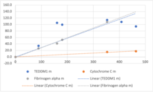
The relative rate of molecular evolution of TEDDM1 was quite similar to that of Fibrinogen Alpha and much more rapid than the evolution rate of Cytochrome C.
Interacting Proteins
Predicted functional partners to the TEDDM1 protein found via STRING.
| Protein | Description | Function |
| SBK2 | Serine/threonine-protein kinase 2 | Predicted to be involved in MAPK cascade and protein phosphorylation |
| METTL7B | Methyltransferase-like protein 7B | Probable methyltransferase |
| PATE2 | Prostate and testis expressed 2 | Contain LY6/PLAUR domain in extracellular space |
| ODF3L1 | Outer dense fiber protein 3-like protein 1 | Active in cytoskeleton |
| DEFB30 | Beta-defensin 130a; Defensin, beta 130 | Antimicrobial host-defense peptide, antiplasmodial activity |
| SPINT3 | Serine peptidase inhibitor, Kunitz type 3 | Enable receptor antagonist activity and beta binding activity |
| TSPAN1 | Tetraspanin-1 | Mediate signal transduction events, role in the regulation of cell development, growth |
| WFDC10A | Wap four-disulfide core domain protein 10a | Functions as a protease inhibitor |
| EDDM3B | Epididymal secretory protein E3-beta | Possible function in sperm maturation |
Clinical Significance
According to previous cancer research studies, gene-based association analyses discovered that TEDDM1 along with GLUL are the two genes most significantly correlated with hepatitis B virus related hepatocellular carcinoma, the most common form of liver cancer. TEDDM1 also has significant clinical significance due to its involvement with keratinocytes and the inhibition of microRNA-31, an important regulator of embryonic implantation and development.
References
- ^ GRCh38: Ensembl release 89: ENSG00000203730 – Ensembl, May 2017
- ^ GRCm38: Ensembl release 89: ENSMUSG00000043282 – Ensembl, May 2017
- "Human PubMed Reference:". National Center for Biotechnology Information, U.S. National Library of Medicine.
- "Mouse PubMed Reference:". National Center for Biotechnology Information, U.S. National Library of Medicine.
- "TEDDM1 transmembrane epididymal protein 1 [Homo sapiens (human)] - Gene - NCBI". www.ncbi.nlm.nih.gov. Retrieved 2022-07-28.
- "GLUL glutamate-ammonia ligase [Homo sapiens (human)] - Gene - NCBI". www.ncbi.nlm.nih.gov. Retrieved 2022-07-28.
- "LINC00272 long intergenic non-protein coding RNA 272 [Homo sapiens (human)] - Gene - NCBI". www.ncbi.nlm.nih.gov. Retrieved 2022-07-28.
- "LOC122149321 Sharpr-MPRA regulatory region 13543 [Homo sapiens (human)] - Gene - NCBI". www.ncbi.nlm.nih.gov. Retrieved 2022-07-28.
- "TEDDM1 Gene - GeneCards | TEDM1 Protein | TEDM1 Antibody". www.genecards.org. Retrieved 2022-07-28.
- "Experiment Detail :: Allen Brain Atlas: Mouse Brain". mouse.brain-map.org. Retrieved 2022-07-30.
- "Services". healthtech.dtu.dk. Retrieved 2022-07-30.
- Attwood MM, Schiöth HB (2021). "Characterization of Five Transmembrane Proteins: With Focus on the Tweety, Sideroflexin, and YIP1 Domain Families". Frontiers in Cell and Developmental Biology. 9: 708754. doi:10.3389/fcell.2021.708754. PMC 8327215. PMID 34350187.
- "PAXdb: Protein Abundance Database". pax-db.org. Retrieved 2022-07-30.
- Novák P, Havlíček V (January 2016). "Protein extraction and precipitation.". Proteomic profiling and analytical chemistry. Elsevier. pp. 51–62. doi:10.1016/B978-0-444-63688-1.00004-5. ISBN 978-0-444-63688-1.
The pI of most proteins is in the pH range of 4 to 7.
- "Compute pI/MW - SIB Swiss Institute of Bioinformatics | Expasy". www.expasy.org. Retrieved 2022-07-28.
- "Motif Scan". myhits.sib.swiss. Retrieved 2022-07-30.
- Kukuruzinska MA, Lennon K (1998). "Protein N-glycosylation: molecular genetics and functional significance". Critical Reviews in Oral Biology and Medicine. 9 (4): 415–448. doi:10.1177/10454411980090040301. PMID 9825220.
- Litchfield DW, Lüscher B (November 1993). Khandelwal RL, Wang JH (eds.). "Casein kinase II in signal transduction and cell cycle regulation". Molecular and Cellular Biochemistry. 127–128. Boston, MA: Springer US: 187–199. doi:10.1007/978-1-4615-2600-1_18. ISBN 978-1-4615-2600-1. PMID 7935350.
- Landschulz WH, Johnson PF, McKnight SL (June 1988). "The leucine zipper: a hypothetical structure common to a new class of DNA binding proteins". Science. 240 (4860): 1759–1764. Bibcode:1988Sci...240.1759L. doi:10.1126/science.3289117. PMID 3289117.
- Bastidas AC, Pierce LC, Walker RC, Johnson DA, Taylor SS (September 2013). "Influence of N-myristylation and ligand binding on the flexibility of the catalytic subunit of protein kinase A". Biochemistry. 52 (37): 6368–6379. doi:10.1021/bi400575k. PMC 3788587. PMID 24003983.
- Lim PS, Sutton CR, Rao S (December 2015). "Protein kinase C in the immune system: from signalling to chromatin regulation". Immunology. 146 (4): 508–522. doi:10.1111/imm.12510. PMC 4693901. PMID 26194700.
- "AlphaFold Protein Structure Database". alphafold.ebi.ac.uk. Retrieved 2022-07-30.
- Rutherford K, Van Duyne GD (2013). "DNA Sequence Recognition by Proteins.". In Lennarz WJ, Lane MD (eds.). Encyclopedia of Biological Chemistry (Second ed.). London: Academic Press. ISBN 978-0-12-378631-9.
The α-helix is the structural element most frequently used for sequence-specific interactions in protein–DNA interfaces. The size of an α-helix matches the width of the DNA major groove, allowing them to fit together tightly while the protein side chains on the helix probe the available base-pair functional groups.
- "TMEM45A transmembrane protein 45A [Homo sapiens (human)] - Gene - NCBI". www.ncbi.nlm.nih.gov. Retrieved 2022-07-28.
- "TMEM45B transmembrane protein 45B [Homo sapiens (human)] - Gene - NCBI". www.ncbi.nlm.nih.gov. Retrieved 2022-07-28.
- "BLAST: Basic Local Alignment Search Tool". blast.ncbi.nlm.nih.gov. Retrieved 2022-07-28.
- "TEDDM1 protein (human) - STRING interaction network". string-db.org. Retrieved 2022-07-28.
- Lin YY, Yu MW, Lin SM, Lee SD, Chen CL, Chen DS, Chen PJ (October 2017). "Genome-wide association analysis identifies a GLUL haplotype for familial hepatitis B virus-related hepatocellular carcinoma". Cancer. 123 (20): 3966–3976. doi:10.1002/cncr.30851. PMID 28662289. S2CID 25961704.
- Stepicheva NA, Song JL (August 2016). "Function and regulation of microRNA-31 in development and disease". Molecular Reproduction and Development. 83 (8): 654–674. doi:10.1002/mrd.22678. PMC 6040227. PMID 27405090.


