| Centroacinar cell | |
|---|---|
 | |
| Details | |
| Location | Pancreas |
| Identifiers | |
| Latin | cellula centroacinosa |
| TH | H3.04.07.0.00008 |
| Anatomical terms of microanatomy[edit on Wikidata] | |
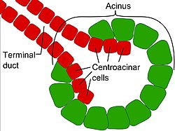
Centroacinar cells are spindle-shaped cells in the exocrine pancreas. They are small and have microvilli on the apical surface. They work with organs such as the kidney, lungs, stomach, brain, intestine.
The exocrine pancreas is one of two compartments that include digestive-acting acinar cells and duct cells.They represent an extension of the intercalated duct into each pancreatic acinus. These cells are commonly known as duct cells, and secrete an aqueous bicarbonate solution under stimulation by the hormone secretin. They also secrete mucin. As well as direct the pathway of the enzyme into the gut, which helps the endocrine pancreas in secreting the hormone into the circulation. This is important in glucose metabolism, insulin-producing B cells, glucagon-producing A cells and somatostatin producing δ cells
The intercalated ducts take the bicarbonate to intralobular ducts which become lobular ducts. These lobular ducts finally converge to form the main pancreatic duct. These intercalated ducts take the bicarbonate to intralobular ducts which become lobular ducts. These lobular ducts finally converge to form the main pancreatic duct. These pancreatic ducts can act as a response mechanism when injury occurs and to avoid disruptions in the pancreatic lumen. This keeps the ionic content in check while keeping it open for flow to occur. Mutations that can occur because of blockage/ accumulation in the pancreatic duct are disorders such as cystic fibrosis.
See also
- List of human cell types derived from the germ layers
- List of distinct cell types in the adult human body
References
![]() This article incorporates text in the public domain from page 1204 of the 20th edition of Gray's Anatomy (1918)
This article incorporates text in the public domain from page 1204 of the 20th edition of Gray's Anatomy (1918)
- ^ "Centroacinar Cells Development in the Pancreatic Duct". LifeMap Discovery. Embryonic.Al. Retrieved May 2, 2024.
- Wendy Lackey M.A. (February 2011). "lectures ession 12". Oral Cavity and Upper GI tract (coursepack from Michigan State University College of Human Medicine). p. 327.
- Beer, Parsons, Roviva, Rebecca L.,Michael J, Meritxell. "Centroacinar cells: At the center of pancreas regeneration". Science Direct. Development Biology. Retrieved May 2, 2024.
{{cite web}}: CS1 maint: multiple names: authors list (link)
External links
- Anatomy Atlases – Microscopic Anatomy, plate 10.213 - "Pancreas"
- Histology image: 10406loa – Histology Learning System at Boston University - "Liver, Gall Bladder, and Pancreas: pancreas, centroacinar cells"
- UIUC Histology Subject 870
| Anatomy of the liver, pancreas and biliary tree | |||||
|---|---|---|---|---|---|
| Liver | |||||
| Biliary tract |
| ||||
| Pancreas |
| ||||
This cell biology article is a stub. You can help Misplaced Pages by expanding it. |