| Transverse fissure of liver | |
|---|---|
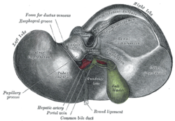
 Inferior surface of the liver. Inferior surface of the liver. | |
 The portal vein and its tributaries. (Porta hepatis labeled at upper left.) The portal vein and its tributaries. (Porta hepatis labeled at upper left.) | |
| Details | |
| Identifiers | |
| Latin | porta hepatis |
| TA98 | A05.8.01.016 |
| TA2 | 3033 |
| FMA | 15758 |
| Anatomical terminology[edit on Wikidata] | |
The porta hepatis or transverse fissure of the liver is a short but deep fissure, about 5 cm long, extending transversely beneath the left portion of the right lobe of the liver, nearer its posterior surface than its anterior border.
It joins nearly at right angles with the left sagittal fossa, and separates the quadrate lobe in front from the caudate lobe and process behind.
Function
It transmits the following (in anterior to posterior order):
- common hepatic duct (leaving)
- proper hepatic artery (entering)
- hepatic portal vein (entering)
The hepatic duct lies in front and to the right, the hepatic artery to the left, and the portal vein behind and between the duct and artery.
It also transmits nerves and lymphatics.
- Sympathetic nerves - these provide afferent pain impulses from the liver and gall bladder to the brain. Pain may be referred to the lower pole of the right scapula (T7).
- Hepatic branch of the vagus nerve (CN X).
Location
The porta hepatis runs in the hepatoduodenal ligament.
When the patient is supine, and the liver observed inferiorly (as in a surgeon's perspective), the important structures demarcating its inferior aspect can be represented by a hepatic "H" figure. The right vertical limb of the "H" defines the left and right functional lobes, while the left vertical limb of the "H" defines the right and left anatomical lobes. The horizontal line between the vertical limbs of the "H" represents the porta hepatis. The quadrate and caudate lobe lie superior and inferior to this line respectively.
See also
References
![]() This article incorporates text in the public domain from page 1191 of the 20th edition of Gray's Anatomy (1918)
This article incorporates text in the public domain from page 1191 of the 20th edition of Gray's Anatomy (1918)
External links
- liver at The Anatomy Lesson by Wesley Norman (Georgetown University)
- Anatomy photo:38:12-0107 at the SUNY Downstate Medical Center - "Stomach, Spleen and Liver: The Visceral Surface of the Liver"
| Anatomy of the liver, pancreas and biliary tree | |||||
|---|---|---|---|---|---|
| Liver | |||||
| Biliary tract |
| ||||
| Pancreas |
| ||||