| Cutaneous myxoma | |
|---|---|
| Other names | Superficial angiomyxoma |
 | |
| Cutaneous myxoma | |
| Specialty | Oncology |
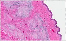
A cutaneous myxoma, or superficial angiomyxoma, consists of a multilobulated myxoid mass containing stellate or spindled fibroblasts with pools of mucin forming cleft-like spaces. There is often a proliferation of blood vessels and an inflammatory infiltrate. Staining is positive for vimentin, negative for cytokeratin and desmin, and variable for CD34, Factor VIIIa, SMA, MSA and S-100.
Clinically, it may present as solitary or multiple flesh-colored nodules on the face, trunk, or extremities. It may occur as part of the Carney complex, and is sometimes the first sign. Local recurrence is common. Cutaneous myxoma is diagnosed based on histopathological features. The differential diagnosis for cutaneous myxoma include alopecia areata, verrucous hamartoma, cyst, fibroma, glioma, hemangioma, lipoma, scar, and nevus sebaceous. Treatment involves complete surgical excision.
Signs and symptoms
Cutaneous myxoma appears as a little, slowly expanding dermal or subcutaneous lump that typically affects adults' heads and necks. Alopecic lesions have been reported, characterized by hair tufts overlaying the lesion or even hypertrichosis. Although usually painless, some people have reported experiencing discomfort or tenderness. A pedunculated lesion was reported.
Causes
Cutaneous myxoma may occur in isolation or in association with syndromes such as nevi, atrial myxoma, myxoid neurofibromas, and ephelides (NAME) syndrome, Carney complex, and lentigines, atrial myxoma, mucocutaneous myxomas, and blue nevi (LAMB) syndrome.
Diagnosis
A thin arborizing vascular network, a large myxoid stroma, mild to moderate cellularity with spindled to stellate cells with little or mild atypia, and the lack of nuclear pleomorphism or mitotic activity are among the histologic markers that are consistently present in cutaneous myxoma.
Alopecia areata, verrucous hamartoma, cyst, fibroma, glioma, hemangioma, lipoma, scar, and nevus sebaceous are only a few of the many conditions included in the extensive clinical differential diagnosis.
Treatment
The main treatment for cutaneous myxoma is complete surgical excision.
See also
References
- Satter EK (October 2009). "Solitary superficial angiomyxoma: an infrequent but distinct soft tissue tumor". J. Cutan. Pathol. 36 (Suppl 1): 56–9. doi:10.1111/j.1600-0560.2008.01216.x. PMID 19187115. S2CID 1528140. Archived from the original on 2012-12-10.
- James, William; Berger, Timothy; Elston, Dirk (2005). Andrews' Diseases of the Skin: Clinical Dermatology (10th ed.). Saunders. p. 614. ISBN 0-7216-2921-0.
- Folpe, Andrew L. (2009). "Soft-Tissue Tumors of the Head and Neck". Diagnostic Surgical Pathology of the Head and Neck. Elsevier. pp. 647–727. doi:10.1016/b978-1-4160-2589-4.00008-5. ISBN 978-1-4160-2589-4.
- Brantsch, K. D.; Metzler, G.; Maennlin, S.; Breuninger, H. (2009). "A meningioma of the scalp that might have developed from a rudimentary meningocele". Clinical and Experimental Dermatology. 34 (8). Oxford University Press (OUP): e792–e794. doi:10.1111/j.1365-2230.2009.03519.x. ISSN 0307-6938. PMID 19817762.
- Miyamoto, Toru; Mihara, Motoyuki; Hagari, Yoshitaka; Shimao, Shuhei (1995). "Primary Cutaneous Meningioma on the Scalp: Report of Two Siblings". The Journal of Dermatology. 22 (8). Wiley: 611–619. doi:10.1111/j.1346-8138.1995.tb03884.x. ISSN 0385-2407. PMID 7560462.
- Peñas, Pablo F. (1995-06-01). "Cutaneous Heterotopic Meningeal Nodules". Archives of Dermatology. 131 (6). American Medical Association (AMA): 731. doi:10.1001/archderm.1995.01690180111022. ISSN 0003-987X. PMID 7778931.
- DA, Lopez; DN, Silvers; EB, Helwig (1974). "Cutaneous meningiomas--a clinicopathologic study". Cancer. 34 (3): 728–744. doi:10.1002/1097-0142(197409)34:3<728::aid-cncr2820340332>3.0.co;2-u. ISSN 0008-543X. PMID 4851231. Retrieved 2024-04-26.
- Ragoowansi, Raj; Thomas, Valerie; Powell, Barry (1998). "Cutaneous meningioma of the scalp: a case report and review of literature". British Journal of Plastic Surgery. 51 (5). Elsevier BV: 402–404. doi:10.1054/bjps.1997.0204. ISSN 0007-1226. PMID 9771369.
- GELLI, M.C.; PASQUINELLI, G.; MARTINELLI, G.; GARDINI, G. (1993). "Cutaneous meningioma: histochemical, immunohistochemical and ultrastructural investigation". Histopathology. 23 (6). Wiley: 576–578. doi:10.1111/j.1365-2559.1993.tb01248.x. ISSN 0309-0167. PMID 7508881.
- ^ Sharma, Anurag; Khaitan, Neha; Ko, Jennifer S.; Bergfeld, Wilma F.; Piliang, Melissa; Rubin, Brian P.; Goldblum, John R.; Billings, Steven D. (2022). "A clinicopathologic analysis of 54 cases of cutaneous myxoma". Human Pathology. 120. Elsevier BV: 71–76. doi:10.1016/j.humpath.2021.12.003. ISSN 0046-8177. PMID 34968506.
- Miedema, Jayson R.; Zedek, Daniel (2012-02-01). "Cutaneous Meningioma". Archives of Pathology & Laboratory Medicine. 136 (2). Archives of Pathology and Laboratory Medicine: 208–211. doi:10.5858/arpa.2010-0505-rs. ISSN 0003-9985. PMID 22288971.
- Ouazzani, A.; de Fontaine, S.; Berthe, J.-V. (2007). "Extracranial meningioma and pregnancy: a rare diagnosis". Journal of Plastic, Reconstructive & Aesthetic Surgery. 60 (6). Elsevier BV: 622–625. doi:10.1016/j.bjps.2006.01.054. ISSN 1748-6815. PMID 17485048.
Further reading
- Iida, Ken; Egi, Takeshi; Shigi, Masato; Sogabe, Yusuke; Ohashi, Hirotsugu (2019). "Cutaneous Myxoma of Multiple Lesions". Plastic and Reconstructive Surgery - Global Open. 7 (2). Ovid Technologies (Wolters Kluwer Health): e2040. doi:10.1097/gox.0000000000002040. ISSN 2169-7574. PMC 6416110. PMID 30881822.
- Alaiti, Samer; Nelson, Fern P.; Ryoo, Jei W. (2000). "Solitary cutaneous myxoma". Journal of the American Academy of Dermatology. 43 (2). Elsevier BV: 377–379. doi:10.1067/mjd.2000.101878. ISSN 0190-9622. PMID 10901728.
External links
| Classification | D |
|---|---|
| External resources |
|