| Thecoma | |
|---|---|
 | |
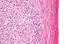
| High magnification micrograph of a thecoma. H&E stain. | |
| Specialty | Oncology |
Thecomas or theca cell tumors are benign ovarian neoplasms composed only of theca cells. Histogenetically they are classified as sex cord-stromal tumours.
They are typically estrogen-producing and they occur in older women (mean age 59; 84% after menopause). (They can, however, appear before menopause.)
60% of patients present with abnormal uterine bleeding, and 20% have endometrial carcinoma.
Pathologic features

Grossly, the tumour is solid and yellow.
Grossly and microscopically, it consists of the ovarian cortex.
Microscopically, the tumour cells have abundant lipid-filled cytoplasm.
References
- Okada I, Nakagawa S, Takemura Y, et al. (October 2004). "Ovarian thecoma associated in the first trimester of pregnancy". J. Obstet. Gynaecol. Res. 30 (5): 368–71. doi:10.1111/j.1447-0756.2004.00212.x. PMID 15327450. S2CID 28580115.
External links
| Classification | D |
|---|
This oncology article is a stub. You can help Misplaced Pages by expanding it. |